Corpus: Scapula


Synonyms: shoulder blade, omoplate
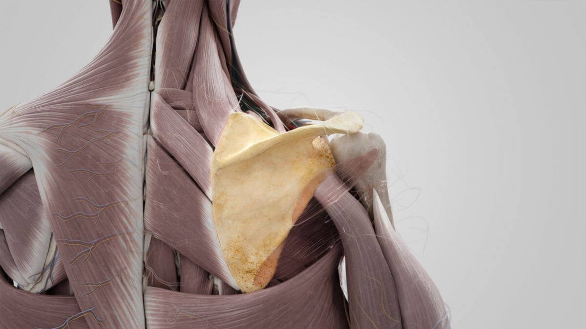
1. Definition
2. Areas
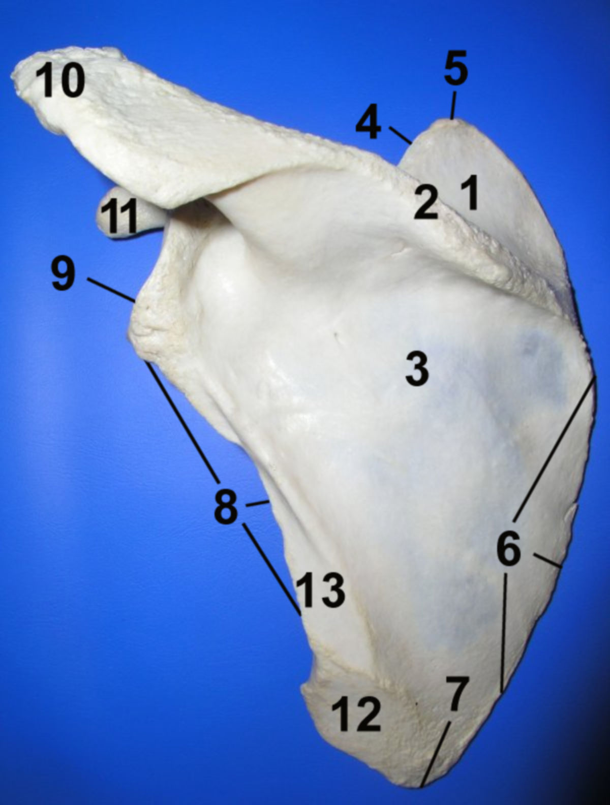
2.1. Dorsal surface
- Divided by the spine of the scapula into the smaller supraspinous fossa and the larger infraspinous fossa.
- The supraspinous fossa is the origin site for the supraspinatus muscle.
- The infraspinous fossa, located below the spine, is predominantly covered by the infraspinatus muscle, originating from its medial two-thirds.
- Laterally, separated by a muscular septum, are the origins for the teres major and teres minor muscles.
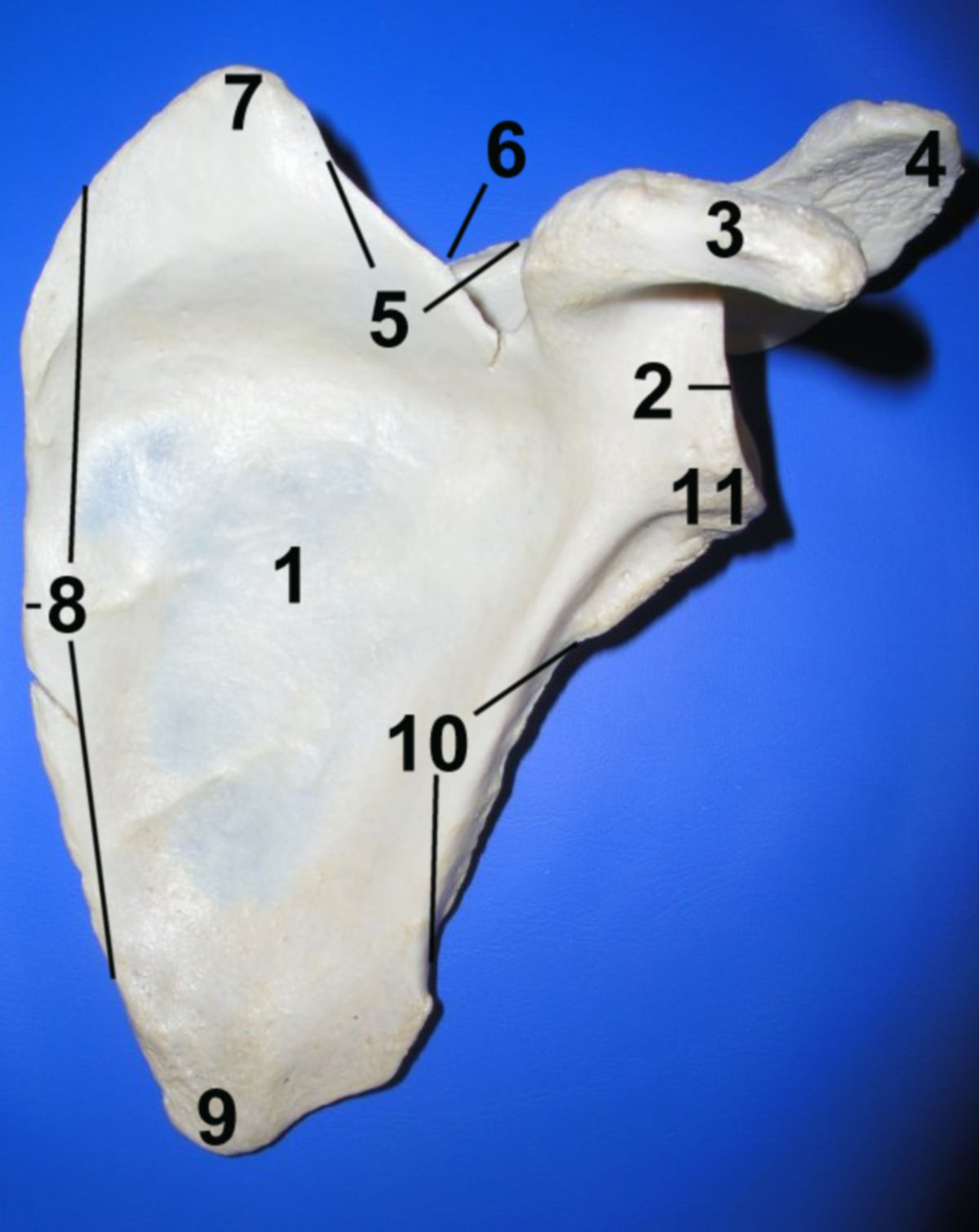
2.2. Ventral surface
- Features an extensive concave subscapular fossa facing the ribs.
- The medial two-thirds of the fossa have oblique edges, serving as the tendon insertions for the subscapularis muscle.
- The subscapular fossa is bordered by smooth triangular areas at the inferior and superior angles, sometimes referred to as the medial angle, where the serratus anterior muscle attaches.
3. Margins
3.1. Superior margin
The shortest edge, extending from the superior angle to the base of the coracoid process. Features the suprascapular notch, bridged by the superior transverse scapular ligament, through which the suprascapular nerve passes. Adjacent areas serve as the insertion point for the omohyoid muscle.
3.2. Lateral margin
The most robust of the three edges, stretching from the lower edge of the glenoid cavity to the inferior angle. Below the glenoid cavity is the infraglenoid tubercle, the origin of the triceps brachii muscle's long head.
3.3. Medial margin
The medial margin is the longest edge, running from the superior to the inferior angle. It serves as the attachment site for several muscles, including the ]]coprus:rhomboid major muscle|rhomboid major]] and minor, the inferior part of the serratus anterior muscle, and the levator scapulae.
4. Angles
4.1. Superior angle
The superior angle is formed by the meeting of the medial and superior margins. It is thin, smooth, and rounded. It is the attachment point for some fibers of the levator scapulae muscle.
4.2. Inferior angle
The inferior angle arises from the convergence of the medial and lateral margins, thick and rough. Dorsal surface serves as the attachment for the teres major muscle and some fibers of the latissimus dorsi muscle.
4.3. Lateral angle
The lateral angle is the most robust part, also known as the "shoulder head." It houses the glenoid cavity, a flat, cartilage-covered socket articulating with the humeral head.
5. Prominent structures
5.1. Spine of scapula
The spine of scapula is a pronounced bony ridge across the dorsal surface, dividing it into the supraspinous and infraspinous fossae. It begins flat at the medial margin, rising laterally and ending in the acromion, which shelters the shoulder joint. The spine of scapula is the attachment site for the trapezius muscle and the origin of the deltoid muscle.
5.2. Coracoid process
The coracoid process is a strong, hook-shaped projection originating near the glenoid cavity. It’s the attachment site for the biceps muscle's short head, the coracobrachialis muscle, the pectoralis minor muscle, and various ligaments, e.g. the coracoacromial ligament and the Coracoclavicular ligament.
5.3. Acromion
The acromion extends from the spine of the scapula, forming the highest point of the clavicle. Its roughened superior surface and lateral edge serve as the origin for the deltoid muscle. Medially, it has a small oval area forming the acromioclavicular joint.
5.4. Glenoid cavity
The glenoid cavity is an oval joint socket with a larger vertical than horizontal diameter, paired with the humeral head to form the shoulder joint. It’s encircled by the glenoid labrum, which deepens the socket. The supraglenoid tuberosity (above) and infraglenoid tuberosity (below) serve as origins for the long heads of the biceps brachii and triceps brachii muscles, respectively.
6. Development
The scapula develops from several ossification centers. During the third fetal month, a large nucleus forms in the areas of the supraspinous and infraspinous fossae and the spine. In the first year, a nucleus appears in the coracoid process, with various smaller nuclei developing across the scapula between the 11th and 18th years. These nuclei typically fuse between the ages of 16 and 22. Occasionally, a nucleus emerging between 15 and 18 years may remain independent.
7. Function
The scapula increases the range of motion of the shoulder girdle and thus the arm through its mobility. Its movements include:
- Vertical translation with movement from caudal to cranial
- Horizontal translation with movement from dorsomedial to ventrolateral
- Rotation with the turning of the inferior angle laterally during abduction or elevation of the arm
The structure of the scapula is a frame construction: The bone tracks parallel to the edge converge in the neck piece (Collum scapulae), which carries the joint socket for the humeral head. This results in the joint pressure being transferred to the frame, mainly absorbed by the lateral margin.
8. Clinic
Paralysis of certain back or shoulder muscles can lead to a protrusion of the scapula, known as scapula alata.