Corpus: Thorax

Synonyms: chest, thoracic cage, rib cage
1. Definition
2. Anatomy
2.1. Overview
The thorax forms the basis for the human chest (pectus) and a large part of the back (dorsum). It consists mainly of hard tissue that protects the thoracic cavity and the upper part of the abdominal cavity as well as the organs contained within them from injury. The thoracic cavity is a cavity divided sagittally into two halves by the mediastinum. The thorax has two openings at the top and bottom:
- the apertura thoracis superior and
- the inferior thoracic aperture, which is closed by the diaphragm.
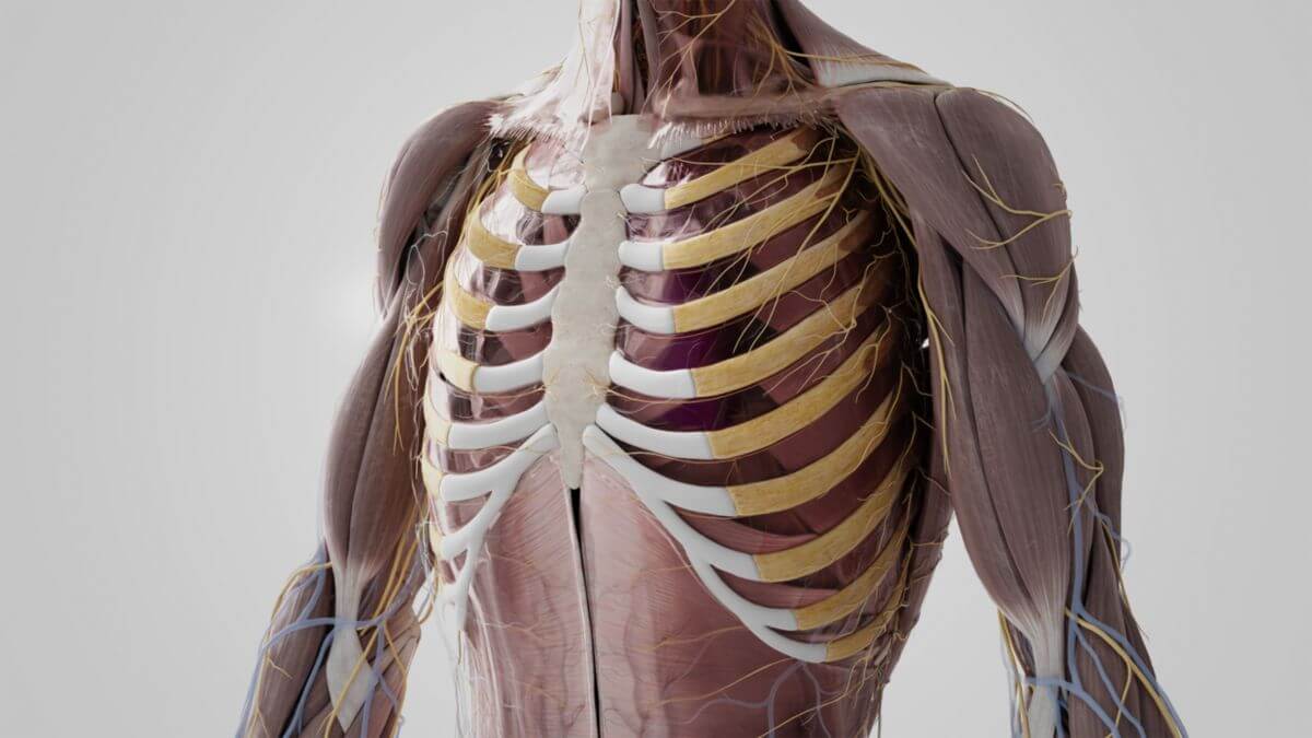
2.2. Thoracic wall
The wall of the thorax is mainly formed by the 12 parallel pairs of ribs, which consist partly of bone and partly of cartilage. The inner and outer intercostal muscles, which are offset like scissors, run between them. On the outside, the thoracic wall is covered by fascia, subcutaneous tissue and skin and partly by muscles. The endothoracic fascia and the parietal leaf of the pleura lie on the inside.
2.3. Contents
The following organs and structures are located within the thorax ("intrathoracic"):
- Chest organs
- Blood and lymph vessels
- Aorta and its major vascular trunks
- Pulmonary artery
- Internal thoracic artery
- Superior vena cava
- Inferior vena cava
- Pulmonary vein
- Internal thoracic vein
- Azygos vein
- Hemiazygos vein
- Thoracic duct
- Nerves
- Ganglia
3. Physiology
The thorax serves to shape the thoracic cavity and as an attachment point for the respiratory muscles. It is an abutment for the capillary forces in the pleural cavity, which guarantee the unfolding of the lungs. This makes the thorax an indispensable morphological component of respiration. During respiration, the thoracic cavity expands in all three anatomical planes, i.e. longitudinally, transversely and sagittally. The longitudinal expansion is primarily caused by the contraction of the diaphragm, which flattens the dome of the diaphragm protruding into the chest and pushes the abdominal organs downwards. The lifting of the ribs is responsible for the sagittal and transverse expansion. Due to their oblique direction, the thoracic volume is significantly increased when the ribs are raised (contraction of the intercostal muscles). The thoracic circumference increases by around 3 to 5 cm during inspiration. Due to adhesion to the parietal pleura, the lungs passively follow the respective change in shape.
4. Malformations
Malformations of the thorax are referred to as thoracic deformities. They can be congenital or acquired and are usually named according to their clinical aspect. Examples are
- Keel chest
- Funnel chest
- Bell-shaped thorax
In addition, there are functional shape adaptations such as the barrel thorax. Deformities of the thoracic spine (e.g. scoliosis) also usually result in thoracic asymmetries, e.g. a rib hump or a Harrenstein deformity. Incomplete thoracic closure in the embryo is called thoracoschisis.
5. Clinic
Due to the location of the central cardiovascular organs (heart, lungs), the thorax is one of the most important areas of the body in medicine, which is primarily dealt with by cardiology and pulmonology. Due to the anatomical complexity of the region, there is a separate branch of surgery for operations on the thorax, thoracic surgery.
5.1. Diagnostics
Various clinical and instrumental methods are available for the examination of the thorax, including
- Percussion
- Auscultation
- Imaging: chest X-ray, chest CT, chest MRI, lung sonography, lung scintigraphy
- Endoscopy: thoracoscopy, mediastinoscopy
- Electrocardiography (ECG)
5.1.1. CT-Fallbeispiel
DICOM-Modelle können auf Mobilgeräten leider nicht angezeigt werden.
5.2. Disease patterns
Important clinical pictures in the area of the thorax are, for example:
- Cardiac infarction
- Pneumonia
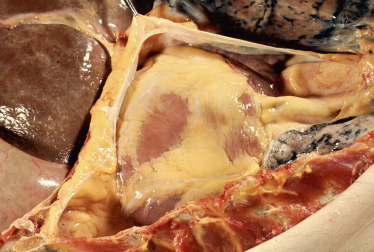
- haematothorax
- Pneumothorax
- Chylothorax
- Tension pneumothorax
- Rib fracture
5.3. Interventions
Access to the thoracic cavity can be achieved by minimally invasive (thoracoscopy) or open surgery (thoracotomy). Accumulations of fluid or gas are removed using a thoracic drain.
6. Source
- 3D model: Dr Claudia Krebs (Faculty Lead) University of British Columbia