Corpus: Auricle


Synonym: pinna
1. Definition
2. Anatomy
The auricle is connected to the periosteum of the skull, also called the pericranium. Its shape is defined by the auricular cartilage, which is a single piece of elastic cartilage.
2.1. Topography
The auricle is located on both sides of the head in the region referred to as the auricular area. It serves as a key reference point for anatomical descriptions. Structures situated in front of the auricle are called preauricular, while those behind it are called retroauricular.
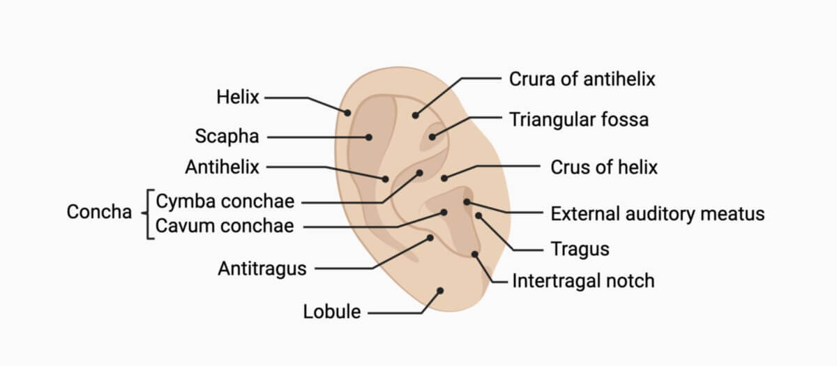
2.2. Morphology
The cartilage of the auricle is highly folded, creating a characteristic relief with multiple ridges and depressions, each of which has a specific name. The outer rim is called the helix. Just inside the helix is a curved indentation called the scapha, which runs parallel to the helix. The next prominent ridge inward is the anthelix, which splits into two folds at its upper end: the superior and inferior roots of the anthelix. Between these roots lies a triangular depression called the triangular fossa.
The anthelix surrounds the concha, a deep hollow that represents the main central portion of the auricle. The concha is divided into two parts by an extension of the helix (the crus helicis): the upper cymba conchae and the lower cavum conchae, which leads into the external auditory canal. Two small protrusions are located near the concha. The tragus, a small anterior projection, is situated closer to the face, while the antitragus is located posteriorly. Between them lies a notch called the intertragic notch. Below the tragus is the earlobe, or lobule, which lacks cartilage and is composed of fatty tissue.
Another feature, Darwin's tubercle, is a small triangular bump on the helix. It is considered an atavistic remnant, representing the tip of a pointed mammalian ear.
The medial surface of the auricle is much less intricate than the lateral surface. Features such as the concha, triangular fossa, and scapha appear as protrusions on this side.
2.3. Ligaments
The auricle is anchored to the skull by three ligaments:
- Anterior auricular ligament: connects the spine of the helix to the root of the zygomatic process of the temporal bone
- Superior auricular ligament: connects the upper edge of the cartilage of the external auditory canal to the squamous part of the temporal bone
- Posterior auricular ligament: connects the back of the concha to the mastoid process of the temporal bone
2.4. Muscles
The auricle's muscles are divided into intrinsic and extrinsic groups.
- Intrinsic muscles: These small, striated muscles connect and stabilize parts of the auricle itself. They include the helicis major and minor, the tragicus, and the antitragicus muscles on the lateral side, and the transverse and oblique auricular muscles on the medial side.
- Extrinsic muscles: These muscles can slightly move the auricle and are part of the facial (mimic) muscles. The anterior and superior auricular muscles attach to the spine of the helix, while the posterior auricular muscle attaches to the eminence of the concha on the medial side. The anterior and superior muscles originate from the scalp, while the posterior muscle originates from the mastoid region.
The auricular muscles are innervated by the facial nerve. The temporal branches of the facial nerve supply the lateral side, while the posterior auricular nerve supplies the medial side.
2.5. Innervation
The sensory innervation of the auricle is provided by several nerves. The medial side is supplied by the cervical plexus:
- Greater auricular nerve (lower two-thirds)
- Lesser occipital nerve (upper third)
The lateral side is innervated by:
- Greater auricular nerve (posterior sections)
- Auricular branches of the vagus nerve (concha and cymba)
- Auriculotemporal nerve from the mandibular nerve of the trigeminal nerve (anterior sections of the helix, tragus, and earlobe)
- Posterior auricular nerve from the facial nerve (ring-shaped area around the concha)
- Communicating branch with the vagus nerve from the glossopharyngeal nerve (entrance to the external auditory canal)
2.6. Blood supply
The medial surface of the auricle is supplied by the auricular branch of the posterior auricular artery, which originates from the external carotid artery or occipital artery. The lateral surface receives blood through branches that perforate the cartilage and by anterior auricular branches of the superficial temporal artery.
2.7. Lymph drainage
Lymphatic drainage from the auricle flows to:
- parotid lymph nodes (anterior sections)
- deep cervical lymph nodes and occipital lymph nodes (remaining sections)
2.8. Histology
The auricular cartilage is composed of elastic cartilage with some fibrocartilage components. It is covered by multilayered keratinized squamous epithelium. The dermis is fused with the perichondrium, and subcutaneous fat is largely absent. The earlobe, however, contains abundant fatty tissue and coarse connective tissue strands.
The skin of the auricle contains lanugo hairs, sebaceous glands, and sweat glands, particularly in the concha and scapha. Longer hairs (tragi hair) are found near the opening of the external auditory canal. Terminal hairs may develop on the tragus and antitragus with age, sometimes forming tufts known as "goat's beard" (hirci barbula).
3. Physiology
The folds and depressions of the auricle serve as a natural filter for incoming sound. These structures refract sound waves based on their frequency, which helps the brain determine the direction of the sound source, particularly whether it is coming from above, below, in front, or behind (spatial hearing).
4. Pathology
Trauma to the auricle (e.g. injuries, insect bites, piercings, frostbite, or surgical interventions) can lead to conditions like auricular hematoma or inflammation of the cartilage (auricular perichondritis). Another common condition is the so called chondrodermatitis nodularis helicis, a painful nodule on the ear's helix or antihelix.
5. Deformities
- Ear cysts
- Ear tags
- Auricular fistulas
- Auricular dysplasia
- Microtia (underdeveloped auricle)
- Prominent ears (often due to excessive curvature of the concha or insufficient angulation of the anthelix)