Corpus: Quadriceps femoris muscle: Unterschied zwischen den Versionen
K (Schützte „Corpus:Quadriceps femoris muscle“ ([Bearbeiten=Nur Administratoren erlauben] (unbeschränkt) [Verschieben=Nur Administratoren erlauben] (unbeschränkt))) |
KKeine Bearbeitungszusammenfassung |
||
| Zeile 1: | Zeile 1: | ||
''Synonym: quadriceps muscle of thigh'' | ''Synonym: quadriceps muscle of thigh'' | ||
== Definition == | == Definition == | ||
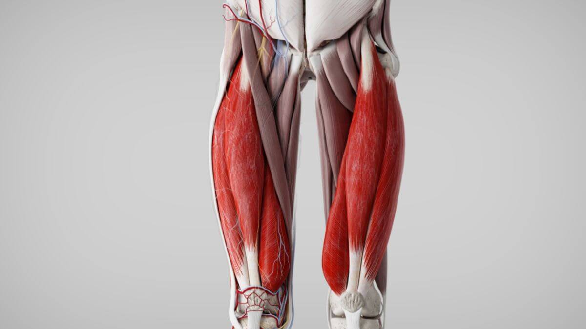
The '''quadriceps femoris muscle''' is located on the ventral side of the thigh | The '''quadriceps femoris muscle''' is located on the ventral side of the [[Corpus:Thigh|thigh]], above the [[Corpus:Articularis genus muscle|articularis genus muscle]]. It consists of four parts: the [[Corpus:Rectus femoris muscle|rectus femoris]], [[Corpus:Vastus intermedius muscle|vastus intermedius]], [[Corpus:Vastus medialis muscle|vastus medialis]], and [[Corpus:Vastus lateralis muscle|vastus lateralis muscles]].<dcembed ratio="16x9"><dcEmbedUrl src="https://www.doccheck.com/de/detail/photos/43219-musculus-quadriceps-femoris"></dcEmbedUrl></dcembed> | ||
== Course == | == Course == | ||
=== | === Rectus femoris muscle === | ||
''from Latin: rectus - right, upright'' | ''from Latin: rectus - right, upright'' | ||
The rectus femoris muscle has two | The rectus femoris muscle has two heads: the straight head which originates from the anterior inferior iliac spine, and the reflected head, a highly variable structure that originates from the upper edge of the [[Corpus:Acetabulum|acetabulum]]. The muscle’s [[Corpus:Tendon|tendon]] begins approximately ten centimeters above the [[Corpus:Patella|patella]]. As a bi-articular muscle, the rectus femoris contributes to both [[Corpus:Knee|knee]] extension and [[Corpus:Hip bone|hip]] flexion. | ||
=== | === Vastus intermedius muscle === | ||
''from Latin: vastus - wide'' | ''from Latin: vastus - wide'' | ||
The vastus intermedius | The vastus intermedius originates from the front and lateral surfaces of the upper two-thirds of the [[Corpus:Femur|femur]]. About halfway down the femur, the muscle fibers converge into a tendon. This muscle is generally located beneath the rectus femoris. | ||
=== | === Vastus lateralis muscle === | ||
The vastus lateralis muscle | The vastus lateralis muscle, the largest of the quadriceps heads, originates mainly from a broad [[Corpus:Aponeurosis|aponeurosis]] attached to the upper part of the intertrochanteric line, the greater trochanter, and the linea aspera of the femur. Some fibers also originate from the lateral intermuscular septum. | ||
=== | === Vastus medialis muscle === | ||
The vastus medialis muscle originates | The vastus medialis muscle originates on the medial side of the femur, from the intertrochanteric line and extending along the medial lip of the linea aspera down to the supracondylar line. | ||
The common tendon of the | The common tendon of the quadriceps muscles attaches to the upper part of the patella. Some fibers extend over the patella, enclosing it as a [[Corpus:Sesamoid bone|sesamoid bone]]. Force is transmitted to the [[Corpus:Lower leg|lower leg]] through the [[Corpus:Patellar ligament|patellar ligament]], which inserts at the tibial tuberosity.<dcembed><dcembedurlskatchfab src="https://sketchfab.com/3d-models/ec745f7384424b8ba24e719a941bbbd1"></dcembedurlskatchfab><dcembedurlskatchfab src="https://sketchfab.com/3d-models/70befb43868f4e1dab5a916e67101f8a"></dcembedurlskatchfab><dcembedurlskatchfab src="https://sketchfab.com/3d-models/3094c775712e4a7f8c53816bfa6fd60d"></dcembedurlskatchfab><dcembedurlskatchfab src="https://sketchfab.com/3d-models/0751006659d94f8ab7570f244be4a894"></dcembedurlskatchfab><dcembedurlskatchfab src="https://sketchfab.com/3d-models/0f46cee03c784739b4152d940dccf033"></dcembedurlskatchfab></dcembed> | ||
<dcembed><dcembedurlskatchfab src="https://sketchfab.com/3d-models/ec745f7384424b8ba24e719a941bbbd1"></dcembedurlskatchfab><dcembedurlskatchfab src="https://sketchfab.com/3d-models/70befb43868f4e1dab5a916e67101f8a"></dcembedurlskatchfab><dcembedurlskatchfab src="https://sketchfab.com/3d-models/3094c775712e4a7f8c53816bfa6fd60d"></dcembedurlskatchfab><dcembedurlskatchfab src="https://sketchfab.com/3d-models/0751006659d94f8ab7570f244be4a894"></dcembedurlskatchfab><dcembedurlskatchfab src="https://sketchfab.com/3d-models/0f46cee03c784739b4152d940dccf033"></dcembedurlskatchfab></dcembed> | |||
== Innervation == | == Innervation == | ||
The quadriceps femoris muscle is innervated by the [[Corpus:Femoral nerve|femoral nerve]] (L2-L4 segments) from the [[Corpus:Lumbar plexus|lumbar plexus]]. | |||
== Function == | == Function == | ||
The quadriceps femoris muscle is the | The quadriceps femoris muscle is the primary extensor of the [[Corpus:Knee joint|knee joint]], essential for straightening the body (e.g., from a squatting position). It works against gravity and is significantly stronger than the hamstrings. The rectus femoris also flexes the [[Corpus:Hip joint|hip joint]], though its effect is relatively weak. Full knee extension by the quadriceps is only achieved when the hip is extended, as the rectus femoris becomes insufficiently effective when the hip is flexed. | ||
<dcembed ratio="16x9"><dcembedurl src="https://www.doccheck.com/de/detail/videos/3104-musculus-quadriceps"></dcembedurl></dcembed> | The quadriceps also stabilize the patella within its groove. Uneven muscle development in this group can lead to patellar dislocation.<dcembed ratio="16x9"><dcembedurl src="https://www.doccheck.com/de/detail/videos/3104-musculus-quadriceps"></dcembedurl></dcembed> | ||
[[Kategorie:Corpus]] | [[Kategorie:Corpus]] | ||
[[Kategorie:Muscle]] | |||
[[Kategorie:Leg]] | |||
Aktuelle Version vom 7. November 2024, 17:33 Uhr
Synonym: quadriceps muscle of thigh
Definition
The quadriceps femoris muscle is located on the ventral side of the thigh, above the articularis genus muscle. It consists of four parts: the rectus femoris, vastus intermedius, vastus medialis, and vastus lateralis muscles.
Course
Rectus femoris muscle
from Latin: rectus - right, upright
The rectus femoris muscle has two heads: the straight head which originates from the anterior inferior iliac spine, and the reflected head, a highly variable structure that originates from the upper edge of the acetabulum. The muscle’s tendon begins approximately ten centimeters above the patella. As a bi-articular muscle, the rectus femoris contributes to both knee extension and hip flexion.
Vastus intermedius muscle
from Latin: vastus - wide
The vastus intermedius originates from the front and lateral surfaces of the upper two-thirds of the femur. About halfway down the femur, the muscle fibers converge into a tendon. This muscle is generally located beneath the rectus femoris.
Vastus lateralis muscle
The vastus lateralis muscle, the largest of the quadriceps heads, originates mainly from a broad aponeurosis attached to the upper part of the intertrochanteric line, the greater trochanter, and the linea aspera of the femur. Some fibers also originate from the lateral intermuscular septum.
Vastus medialis muscle
The vastus medialis muscle originates on the medial side of the femur, from the intertrochanteric line and extending along the medial lip of the linea aspera down to the supracondylar line.
The common tendon of the quadriceps muscles attaches to the upper part of the patella. Some fibers extend over the patella, enclosing it as a sesamoid bone. Force is transmitted to the lower leg through the patellar ligament, which inserts at the tibial tuberosity.
Innervation
The quadriceps femoris muscle is innervated by the femoral nerve (L2-L4 segments) from the lumbar plexus.
Function
The quadriceps femoris muscle is the primary extensor of the knee joint, essential for straightening the body (e.g., from a squatting position). It works against gravity and is significantly stronger than the hamstrings. The rectus femoris also flexes the hip joint, though its effect is relatively weak. Full knee extension by the quadriceps is only achieved when the hip is extended, as the rectus femoris becomes insufficiently effective when the hip is flexed.
The quadriceps also stabilize the patella within its groove. Uneven muscle development in this group can lead to patellar dislocation.