Corpus: Patella


Synonym: kneecap
1. Definition
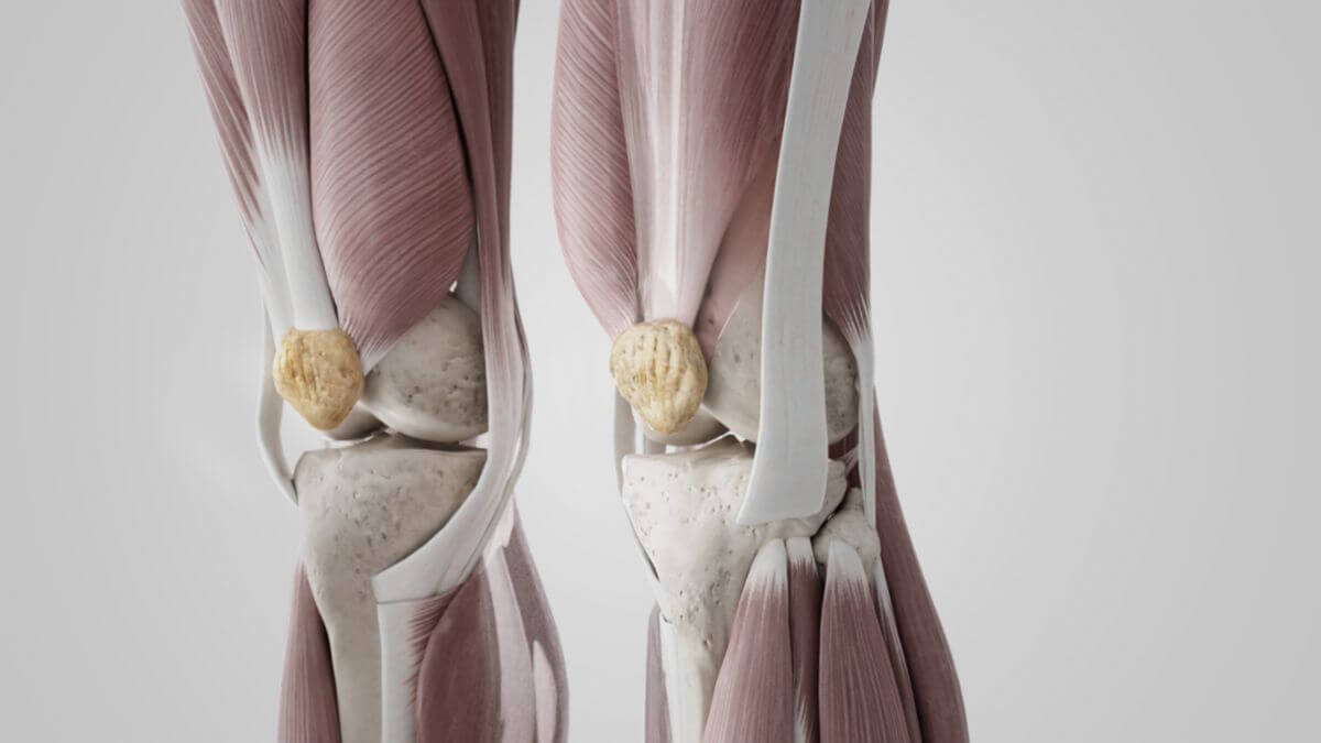
The patella is a flat, disc-shaped bone, triangular when viewed from the front, which is located in front of the knee joint, in whose articular surfaces it is involved. The patella acts as a sesamoid bone in the tendon of the quadriceps femoris muscle. It protects the knee joint and extends the lever arm of the quadriceps femoris muscle.
2. Surfaces
The patella has a concave and convex side due to its adaptation to the femoral condyles.
2.1. Anterior surface
The front of the patella is convex and has small openings through which the supplying vessels pass into the interior of the bone. It is completely covered by the tendon of the quadriceps femoris muscle, which continues caudally into the patellar ligament. Further forwards, a bursa (prepatellar bursa) provides cushioning and mobility in relation to the skin.
2.2. Posterior surface
The concave back of the patella is covered by articular cartilage in the upper two thirds and has a vertical ridge, the crista patellae. It inserts into the intercondylar sulcus between the condyles of the femur. The crista patellae divides the back into a large lateral and a smaller medial joint facet, which in turn articulate with the condyles of the femur. The facets form an angle of 120 to 140° in the ridge (patella opening angle). Furthermore, the so-called Haglund dent is often found in the center of the ridge, which is balanced by thick articular cartilage. The medial facet often has a secondary facet at the medial edge that is angled forwards, the so-called odd facet.
3. Margins
3.1. Superior edge
The upper, strong edge of the patella serves as an insertion surface for the quadriceps femoris muscle, more precisely for two of its muscle heads, the rectus femoris muscle and the vastus intermedius muscle.
3.2. Medial and lateral edges
The medial and lateral edges taper caudally and serve as an attachment surface for the vastus lateralis and vastus medialis muscles.
3.3. Apex patellae
The caudally extending apex of the patella serves as the origin for the patellar ligament, which runs to the tibia.
4. Tendons
The majority of the tendon fibers of the quadriceps femoris muscle converge proximally to the patella and partially cross over each other. These fibers radiate into the upper pole of the patella and are anchored there in the compacta. The medial and lateral portions of the quadriceps tendon run along the sides of the patella directly to the tibial tuberosity. Fibers further radiate into the capsule of the knee joint and form the retinaculum patellae. The distal tendon fibers originate on the anterior surface and at the lower pole (apex patellae), which then run as the patellar ligament to the tibial tuberosity of the tibia.
5. Function
The patella is a hypomochlion for the tendon of the quadriceps femoris muscle. It increases the distance of the force vector of the quadriceps femoris muscle from the center of rotation of the knee joint and thus extends the lever arm of the extensor muscles of the thigh. Thanks to the cartilage surface, the patella reduces the sliding resistance of the patellar tendon. During the extension movement, the patella covers a distance of around 8-10 cm over the femur bone.
In addition, the patella also protects the joint space from the penetration of foreign bodies when force is applied to the knee.
6. Varieties
- Bipartite patella: Patella consists of two separate bone parts
- Multipartite patella: Patella consists of several separate bone parts
- Patella emarginata: Indentation at the proximal edge of the patella
7. Clinic
Diseases of the patella belong to the speciality of orthopaedics and trauma surgery. They include, among others:
- Chondropathia patellae
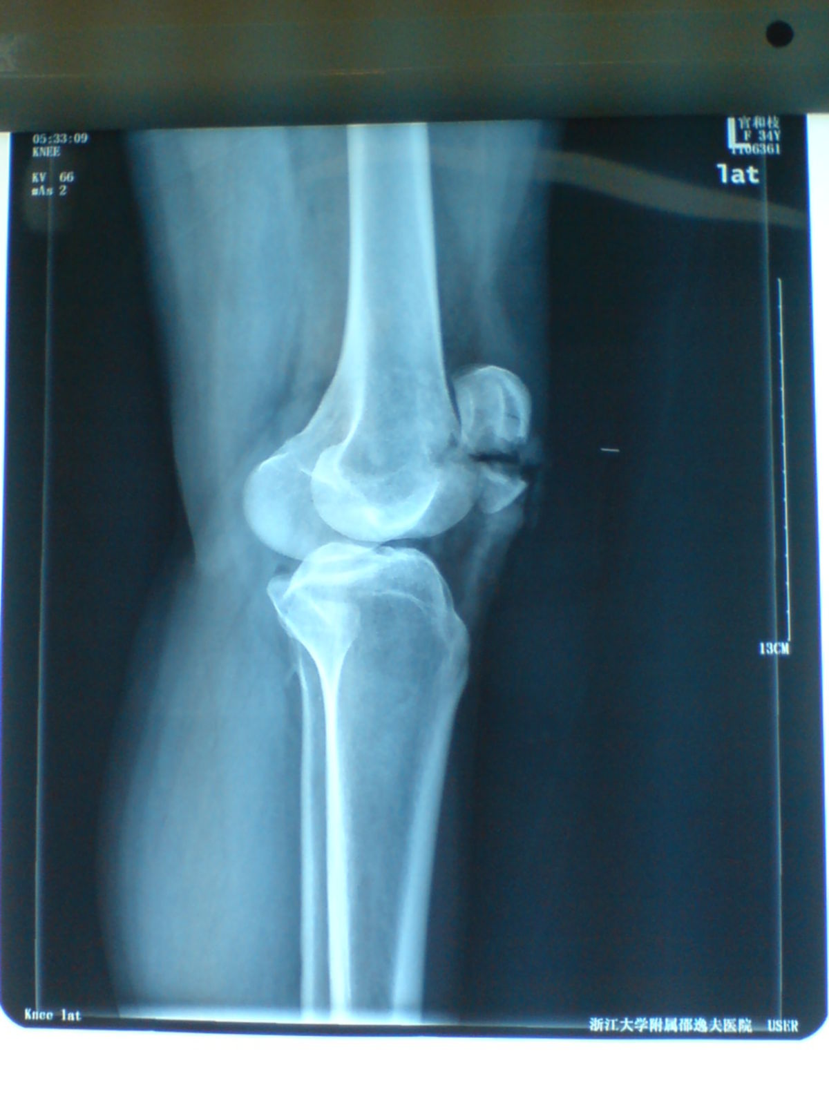
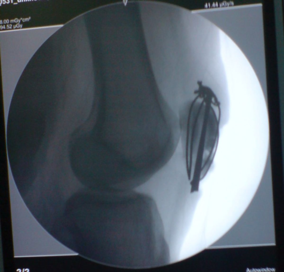
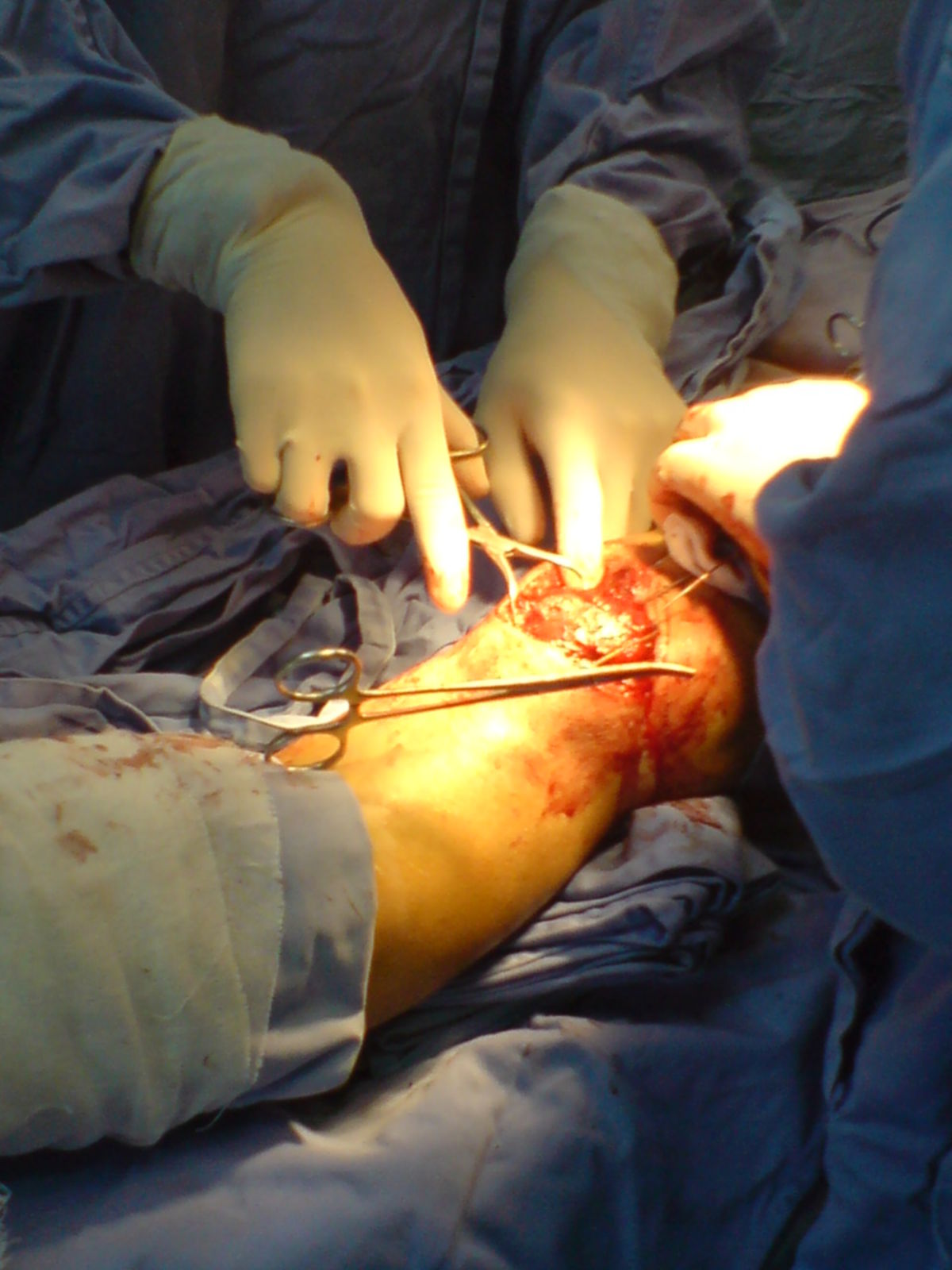
- Patella fracture
- Retropatellar arthrosis
- Patella alta