Corpus: Calcaneus


from Latin: calcar - spur
Synonyms: os calcis, heel bone
1. Definition
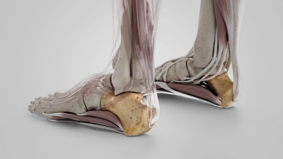
The calcaneus is the largest bone of the foot skeleton. It is involved in the formation of the talotarsal joint and serves as a lever for the flexors of the lower leg, which attach to it via the Achilles tendon.
2. Anatomy
The calcaneus has a cubic shape and shows 6 surfaces.
2.1. Surfaces
2.1.1. Superior surface
The superior surface of the calcaneus is essentially occupied by 3 articular facets that articulate with the talus:
- Posterior talar articular surface
- Middle talar articular surface
- Anterior talar articular surface
The calcaneal sulcus lies between the posterior and medial articular talar surface. Together with the talar sulcus of the talus, it forms the so-called sinus tarsi, a tunnel that accommodates the interosseous talocalcaneal ligament. The posterior talar articular surface is involved in the formation of the posterior talotarsal joint, the anterior and media surfaces in the formation of the anterior talotarsal joint. The small, rough bony facet in front of the articular surfaces serves as the origin of the extensor digitorum brevis muscle.
2.1.2. Inferior surface (plantar surface)
The inferior surface is uneven and wider posterior than in front. It is bordered posteriorly by a prominent elevation, the calcaneal tuber (tuber calcanei). The Achilles tendon is attached to it. The tuberosity forms a small bony process on both sides. The medial process (tuberis calcanei) on the inner side serves as the origin for the abductor hallucis muscle and the flexor digitorum brevis muscle. From its base to the lateral process (tuberis calcanei), which is located on the outside of the foot, the origin of the abductor digiti quinti muscle runs across the bone. The two processes also serve to anchor the plantar aponeurosis. There is a central depression between the two processus. Further anterior to the processus, the two heads of the quadratus plantae muscle are located medially and laterally.
2.1.3. Medial surface
The medial side of the calcaneus is concave in shape. At its cranial end there is a prominent, horizontal bony projection, the sustentaculum tali. The underside is shaped like a groove towards the sulcus tendinis musculi flexoris hallucis longi and accommodates the tendon of the flexor hallucis longus muscle.
2.1.4. Lateral surface
The lateral side of the calcaneus has a slightly protruding bony tubercle (trochlea fibularis). The sulcus tendinis musculi peronei longi, which holds the tendon of the peroneus longus muscle, lies under the trochlea fibularis.
2.1.5. Anterior Surface
The anterior side of the calcaneus has a connecting surface to the cuboid bone, the so-called facies articularis cuboidea.
3. Development
The bony core of the calcaneus develops between the 4th and 7th foetal month.
4. Function
The calcaneus serves as a lever for the most important flexors of the lower leg muscles. Furthermore, through the origin of the plantar aponeurosis, it plays a significant role in maintaining the tension of the arch of the foot.
5. Clinic
Common pathological changes of the calcaneus are the so-called heel spur and Haglund's deformity. These involve ossification of the tendon insertions of the plantar aponeurosis ("plantar heel spur") or the Achilles tendon ("cranial heel spur"). Both variants often remain asymptomatic, but can also lead to pain on exertion. Calcaneus fractures are rather rare due to the compact structure of the bone and occur primarily in cases of massive force, e.g. when falling from a great height. Fatigue fractures of the calcaneus occur in athletes who participate in jumping sports.