Corpus: Gallbladder


1. Definition
The gallbladder serves as a storage organ for 30-80 ml of bile. By removing water, the bile is concentrated, enabling a larger amount to be stored.
2. Anatomy
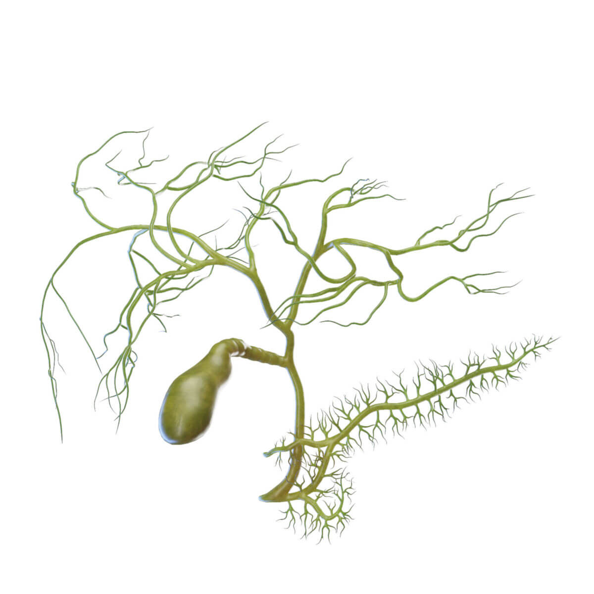
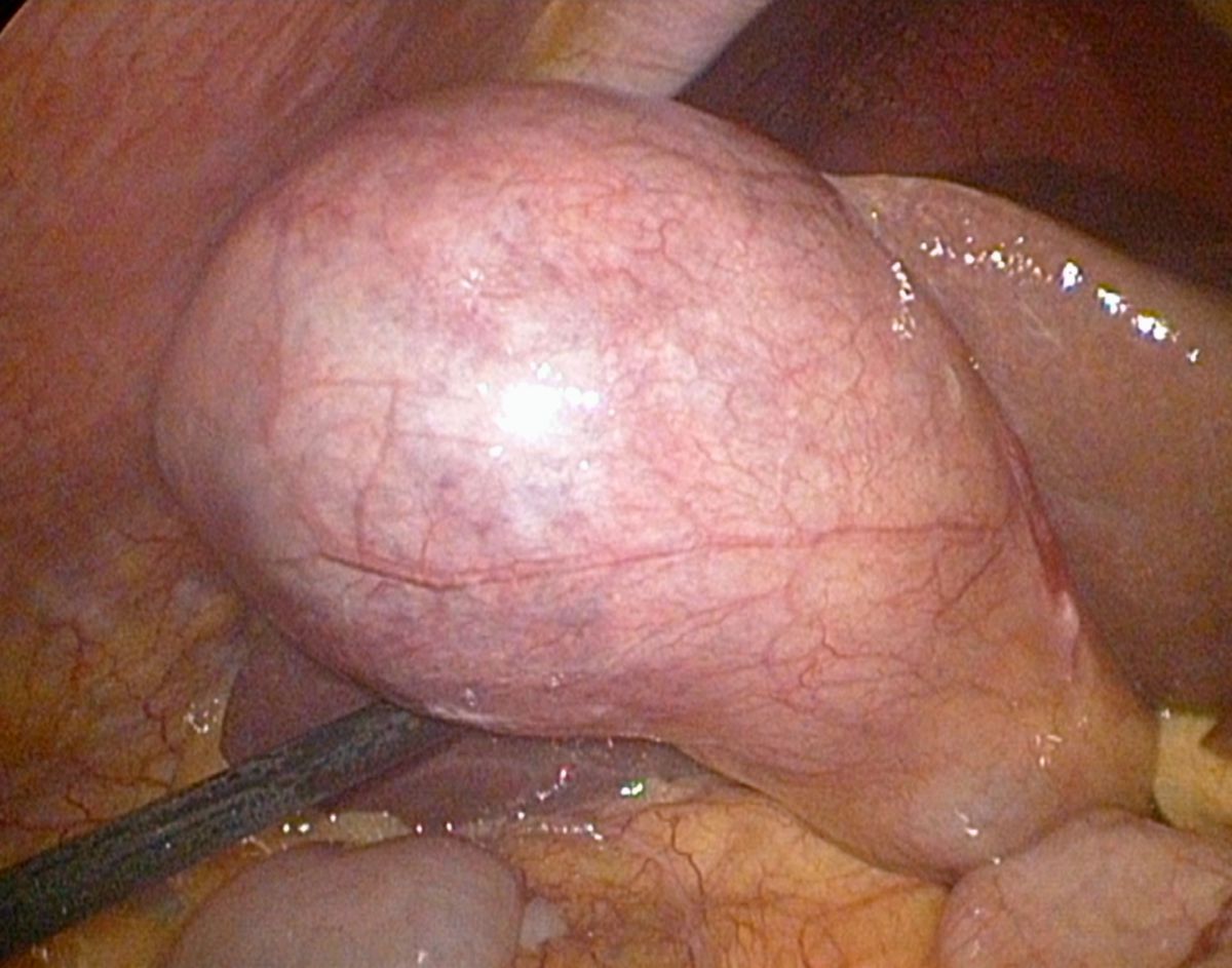
The gallbladder is pear-shaped and located on the underside of the liver in the gallbladder fossa. It is attached to the liver's visceral surface by firm connective tissue that is part of Glisson's capsule. The side facing the intestine is covered by peritoneum. The gallbladder is approximately 8 cm long and 4 to 5 cm wide. Under normal conditions, the wall thickness is less than 4 mm when fasting and less than 8 mm after eating.
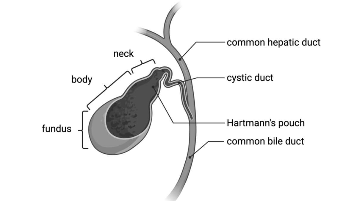
The gallbladder is divided into three anatomical parts:
- Fundus: the bottom of the gallbladder.
- Body: the main portion of the gallbladder.
- Neck: the narrow part that connects to the cystic duct.
The neck of the gallbladder and the cystic duct contain a spiral fold of mucosa called the spiral valve (Heister's valve), which helps prevent bile outflow. In the region of the neck, there is also a small pouch called Hartmann's pouch.
2.1. Vascular supply
The gallbladder receives its blood supply from the cystic artery, which arises from the right branch of the hepatic artery. The veins of the gallbladder drain into the portal vein via the hepatoduodenal ligament.
2.2. Innervation
The gallbladder and bile ducts are innervated by the autonomic nervous system through the hepatic plexus, which originates from the celiac plexus. Autonomic stimulation increases gallbladder contraction and bile duct relaxation, enhancing bile flow. The hepatic plexus also contains afferent fibers that carry pain signals. Additional pain fibers run from the peritoneum covering the gallbladder to the right phrenic nerve, explaining why gallbladder pain can radiate to the right shoulder.
2.3. Palpation
The gallbladder may be palpated when enlarged, located below the liver's edge, medial to the midclavicular line, and can protrude beneath the right costal margin during breathing. Two clinical signs associated with gallbladder palpation are the Murphy's sign (pain with deep inspiration during palpation) and the Courvoisier’s sign (an enlarged, painless gallbladder, often associated with bile duct obstruction)

3. Histology
The gallbladder wall consists of several layers:
- The mucosa is lined with a single layer of columnar epithelium with microvilli that produce mucus to protect the mucosa from bile. The mucosa has prominent folds, which can form crypt-like extensions known as Rokitansky-Aschoff sinuses or Luschka's crypts of the gall bladder. Mucosal bridges are also commonly seen.
- The muscularis layer consists of smooth muscle fibers arranged in a lattice-like pattern, facilitating contraction.
- There is no muscular layer of the mucosa or submucosa.
- The subserosal connective tissue connects to the fibrous capsule of the liver (Glisson's capsule).
- The serosa covers the parts of the gallbladder that are not attached to the liver.
4. Physiology
Bile produced by the liver travels to the duodenum via the common hepatic and common bile ducts. If the sphincter of hepatopancreatic ampulla (sphincter of Oddi) is closed, bile is diverted into the gallbladder via the cystic duct for storage. The gallbladder empties when its muscle wall contracts, a process stimulated by the hormone cholecystokinin, which is secreted by intestinal endocrine cells, and by acetylcholine from parasympathetic fibers of the vagus nerve.
5. Clinic
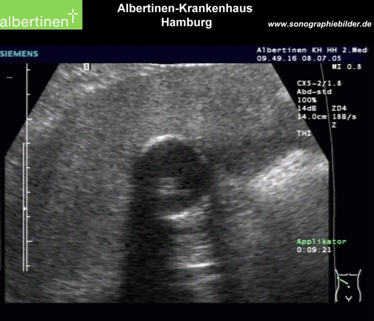
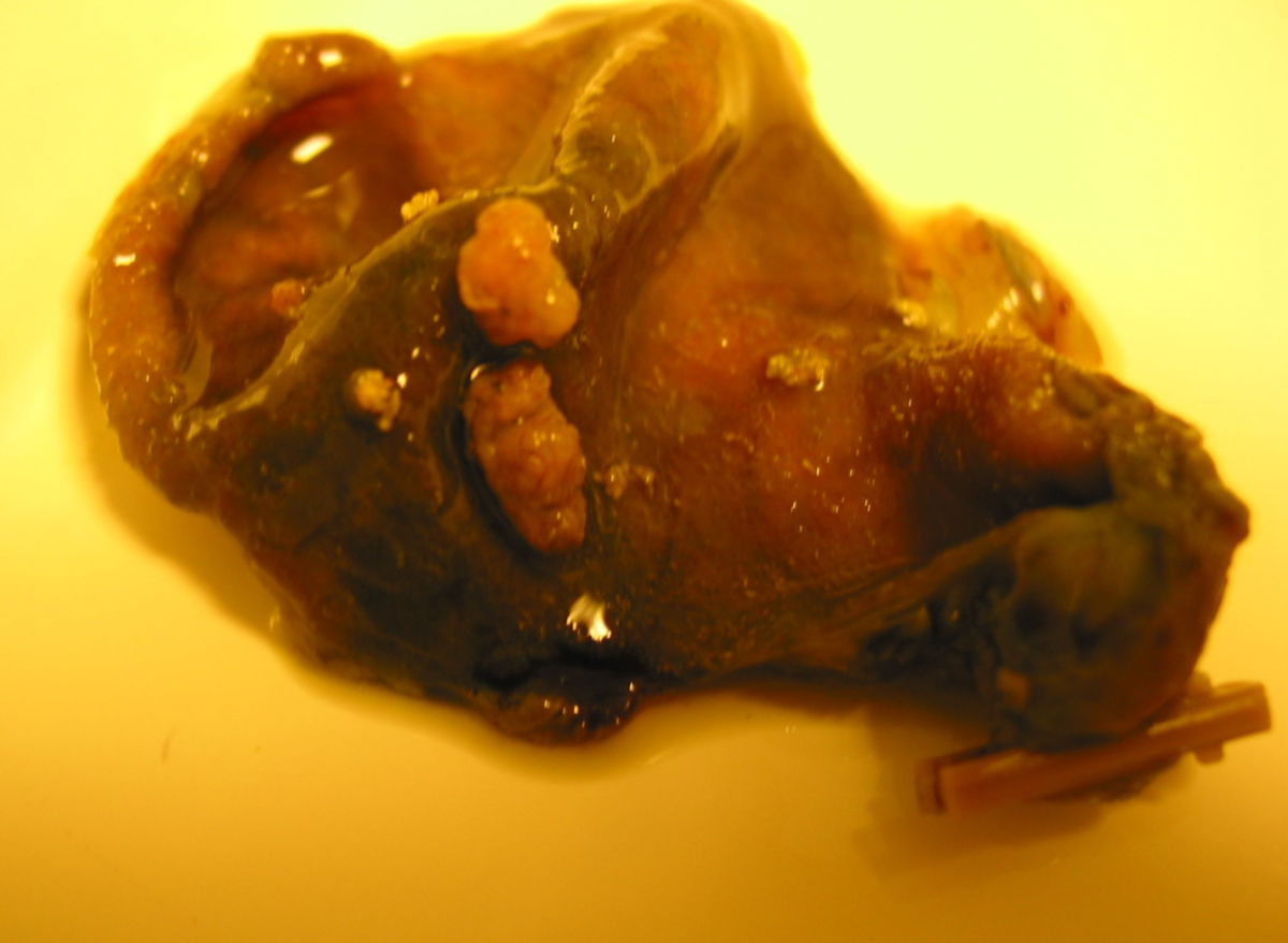
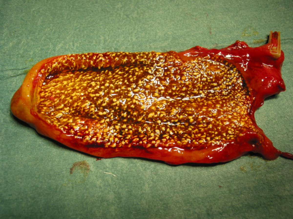
The most common disease of the gallbladder is cholelithiasis, affecting about 15 % of adults. It results from a disturbance in cholesterol metabolism and can cause biliary colic, characterized by intense abdominal pain.
A serious complication is gallbladder perforation, where bile leaks into the abdominal cavity, often resulting in peritonitis and an acute abdomen, a medical emergency.
6. Image source
- Illustration: human gallbladder and biliary tree by www.sciepro.com; CC BY-ND 4.0 EN