Corpus: Liver


1. Definition
The liver is the largest and most essential metabolic organ in the human body. Its functions include processing nutrients from food, breaking down and excreting substances, and producing vital proteins.
2. Overview
The liver is the largest digestive gland in the human body, producing plasma proteins, antibodies, and bile. It serves as the primary detoxification organ. Most nutrients absorbed into the blood from the intestines first pass through the liver, which regulates their release or storage as needed. The liver also stores vitamins and produces precursors for hormone synthesis.
3. Embryonic development
The liver develops from an epithelial bud of the embryonic foregut and differentiates into a mature organ, making it a derivative of the endoderm. The development of the liver can be divided into two stages:
- Development of the liver parenchyma, bile ducts, and gallbladder.
- Development of the intrahepatic vascular system.
4. Anatomy
In adults, the liver weighs about 1,400 to 1,800 g. It is a soft, uniformly structured organ located in the right upper abdomen. Macroscopically, the liver is divided into four lobes:
- Right lobe
- Left lobe
- Quadrate lobe
- Caudate lobe
The right lobe lies beneath the diaphragm and is partially fused to it. It is larger than the left lobe, which extends into the left upper abdomen.
The liver has two surfaces:
- Diaphragmatic surface: Convex and facing the diaphragm.
- Visceral surface: Concave, facing the abdominal organs, with recesses for neighboring structures like the gallbladder.
The liver receives blood from the portal vein, which carries nutrients from the digestive tract, and from the hepatic artery proper. Blood is drained via hepatic veins into the inferior caval vein.
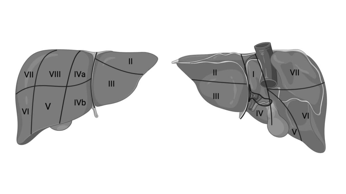
Functionally, the liver is divided into eight segments.
5. Preparation
6. Histology
The liver is composed of 1-2 mm structural units called hepatic lobules. These lobules have a hexagonal cross-section and are organized around a central vein. Each lobule consists of columns of hepatocytes arranged around hepatic sinusoids, which are blood-filled spaces lined with discontinuous endothelium. Specialized macrophages called Kupffer cells are found within the sinusoids.
Between the endothelial cells of the sinusoids and the hepatocytes is the space of Disse, where exchange between the blood and hepatocytes occurs. Ito cells, which store vitamin A and fat, are located here.
The periportal fields lie at the junction of three liver lobules and contain branches of the hepatic artery, portal vein, and intrahepatic bile ducts. These structures form the Glisson triad. Blood flows through the sinusoids from the portal vein and hepatic artery toward the central vein, eventually draining into the hepatic veins and inferior caval vein.
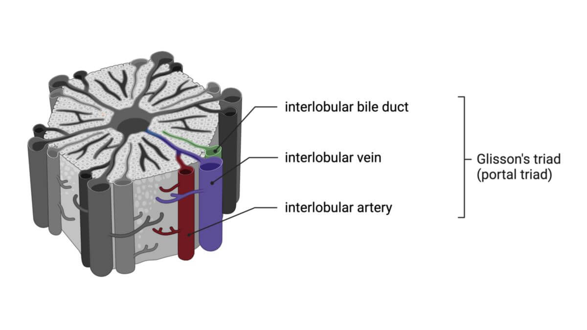
The hepatic sinusoids transport blood from the interlobular vein (derived from the portal vein) and interlobular artery through the hepatic lobules toward the center, where it is collected by the central vein. The central veins then merge to form larger sublobular veins, which ultimately flow into the hepatic veins that empty into the inferior caval vein.
Metabolic products of the hepatocytes, destined for excretion, are secreted into bile canaliculi, small cavities within the hepatocytes. Upon exiting the hepatic lobules, these bile canaliculi converge to form bile ducts, which are lined by a single layer of prismatic epithelium. These ducts combine to form larger bile ducts, eventually leading to the right and left hepatic ducts.
In addition to the anatomical division into hepatic lobules, the liver tissue can also be functionally divided into hepatic acini and portal lobules.
7. Function
7.1. Overview
The liver plays a central role in glucose, fat, and protein metabolism. It regulates blood sugar levels by absorbing glucose from the blood and storing it as glycogen. When needed, glycogen is broken down into glucose. The liver also controls hormone regulation (e.g., insulin), which helps maintain stable blood sugar levels, and influences fat breakdown.
7.2. The tasks in detail
- Storage
- Glucose as glycogen
- Fats as lipoproteins
- Vitamins (A, B12, D, E, K)
- Iron
- Blood
- Protein production
- Albumin
- Transferrin
- Coagulation factors
- Antithrombin
- Plasminogen
- Fat and lipoprotein synthesis
- Triacylglycerides
- Phospholipids
- Sphingolipids
- Very-low-density lipoprotein (VLDL)
- Bile formation
- Cholesterol and bile acid synthesis
- Detoxification and waste breakdown
- Ammonia to urea conversion
- Breakdown of red and white blood cells
- Hemoglobin degradation to bilirubin
- Drug metabolism
- Alcohol breakdown (ethanol oxidation)
- Regulation of acid-base balance
- Defense
- Immune defense against pathogens from the gastrointestinal tract
- Blood formation
- Haematopoiesis in the fetus up to the seventh month of pregnancy
8. Clinic
8.1. Important liver diseases
Liver diseases, known as hepatopathies, fall under the field of hepatology, a subspecialty of gastroenterology. Common liver diseases include:
- Fatty liver disease
- Alcoholic hepatitis
- Viral hepatitis (A, B, C, D, E)
- Chronic hepatitis
- Liver cirrhosis
- Liver failure
- Liver metastases
- Hepatocellular carcinoma (HCC)
- Liver abscesses
8.2. Diagnostics
The liver can be examined through various methods, such as:
- Palpation: Feeling for enlargement or tenderness.
- Inspection: Checking for signs like jaundice, ascites, edema, or spider nevi.
- Imaging techniques:
- Ultrasound
- CT scans
- MRI
- Magnetic resonance elastography (MRE)
- Endoscopic retrograde cholangiopancreatography (ERCP)
- Liver biopsy
- Laboratory diagnostics: Measuring liver enzymes and other blood parameters to assess liver function.
8.3. Laboratory diagnostics
8.3.1. Liverenzymes
When liver cells are damaged, specific enzymes increase in the blood. The type and level of enzyme elevation can help determine the nature of the liver disease. Common liver enzymes measured include:
- Alanine aminotransferase (ALT), formerly known as glutamate pyruvate transaminase (GPT).
- Aspartate aminotransferase (AST), formerly known as glutamate oxaloacetate transaminase (GOT).
- Glutamate dehydrogenase (GLDH).
- Gamma-glutamyl transferase (GGT).
- Alkaline phosphatase (AP).
8.3.2. Further laboratory parameters
- Bilirubin (direct and indirect)
- Bilirubin in urine
- Cholinesterase, albumin
- Quick value, antithrombin
- Hepatitis serology
- Serum iron, ferritin, copper, and ceruloplasmin levels.
9. Literature
- "Therapie von Leber- und Gallekrankheiten", ed. by Wolfgang F. Caspary, Ulrich Leuschner and Stefan Zeuzem. Springer Verlag Berlin (2001) ISBN 3-540-67390-3
- "Spezielle pathologische Anatomie, Bd. 10: Pathologie der Leber und Gallenwege", ed. by Gerhard Seifert, by Helmut Denk, H. P. Dienes, Jochen Düllmann. Springer Verlag Berlin (2000) ISBN 3-540-65511-5
- Liver Diseases", ed. by Ellen Schmidt, Friedrich W. Schmidt and Michael P. Manns (2000) ISBN 3-8047-1640-7
- Practical Hepatology", Kuntz, Erwin; Kuntz, Hans-Dieter. Karl F. Haug Fachbuchverlag (1998) ISBN 3-8304-0479-4