Corpus: Fibula


from latin: fibula - clasp
synonym: calf bone
1. Definition
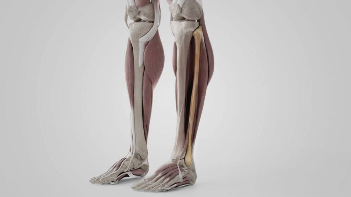
The fibula is a bone of the leg which, together with the tibia, forms the bony lower leg. Firmly attached to the tibia, the fibula forms the articular surface for the talocrural joint.
2. Anatomy
2.1. Corpus fibulae (fibular shaft)
The fibular shaft has three sharp edges (anterior, posterior and interosseous) that grasp three surfaces (medial, lateral and posterior) between them. The multiple division is due to the numerous muscle origins. The tight interosseous membrane extends between the interosseous crest and the edge of the tibia of the same name, dividing the entire lower leg into an anterior and a posterior section. On the posterior surface of the fibular shaft, the crista medialis separates the area of origin of the posterior tibialis muscle from that of the flexor hallucis longus muscle.
2.2. Collum fibulae (fibula neck)
The fibula neck (collum fibulae) is the connection between the head and body of the fibula.
2.3. Caput fibulae (fibula head)
Like the adjoining "neck part" (collum fibulae), the head of the fibula is easy to feel on the lateral side of the knee. Contact with the tibia is made via a cartilaginous articular surface, the facies articularis capitis fibulae, which is connected to the facies articularis fibularis tibiae on the lateral condyle of the tibia. The very prominent tip of the fibula, the "apex capitis fibulae", points proximally.
2.4. Malleolus lateralis (lateral malleolus)
The angular cross-sectional shape of the fibula becomes less pronounced towards the lower end. Here a strong flare forms the lateral malleolus. This lies close to the tibia and has its own articular surface — the facies articularis malleolaris lateralis. The lateral malleolus extends further distally than the tibia. Together with the medial malleolus of the tibia, it forms the malleolar fork, which grasps the talus between them.
3. Development
In the 2nd embryonic month, the perichondral bone cuff develops in the area of the corpus. An enchondral bone nucleus appears in the malleolus in the 2nd year of life, but not in the fibular head until the 4th year of life. Closure of the epiphyses occurs distally between the ages of 16 and 19, proximally somewhat later, between the ages of 17 and 20. The course of the epiphyseal joint line lies proximally below the fibular head, distally slightly above the malleolus.
4. Function
The fibula is not connected to the knee joint, but its distal end forms part of the talocrural joint; from there it transmits the forces acting on the leg via the tibiofibular joint and the middle tibiofibular ligament to the tibia and thus to the femur.