Corpus: Coronary artery: Unterschied zwischen den Versionen
(Die Seite wurde neu angelegt: „{{Raw}} ''from Latin: corona - wreath, crown '' ==Definition== The '''coronary arteries''' are the two arteries that surround the heart in a ring shape and supply the heart muscle with blood - including the branches that branch off from these vessels. <dcembed viewer-ident="yZfd4KeBB36WQfqPjJTwgA"><dcembedurl src="https://www.doccheck.com/en/detail/photos/1343-muscular-bridge-lad"></dcembedurl> <dcembedurl src="https://www.doccheck.com/en/detail/photos…“) |
(Bis Abschnitt "Overview" übersetzt) |
||
| Zeile 4: | Zeile 4: | ||
==Definition== | ==Definition== | ||
The '''coronary arteries''' are the two | The '''coronary arteries''' are the two blood [[Corpus:Vessel|vessels]] that encircle the [[Corpus:Heart|heart]] like a ring and supply blood to the heart [[Corpus:Muscle|muscle]], including their branching vessels. | ||
<dcembed viewer-ident="yZfd4KeBB36WQfqPjJTwgA"><dcembedurl src="https://www.doccheck.com/en/detail/photos/1343-muscular-bridge-lad"></dcembedurl> | <dcembed viewer-ident="yZfd4KeBB36WQfqPjJTwgA"><dcembedurl src="https://www.doccheck.com/en/detail/photos/1343-muscular-bridge-lad"></dcembedurl> | ||
| Zeile 14: | Zeile 14: | ||
==Anatomy== | ==Anatomy== | ||
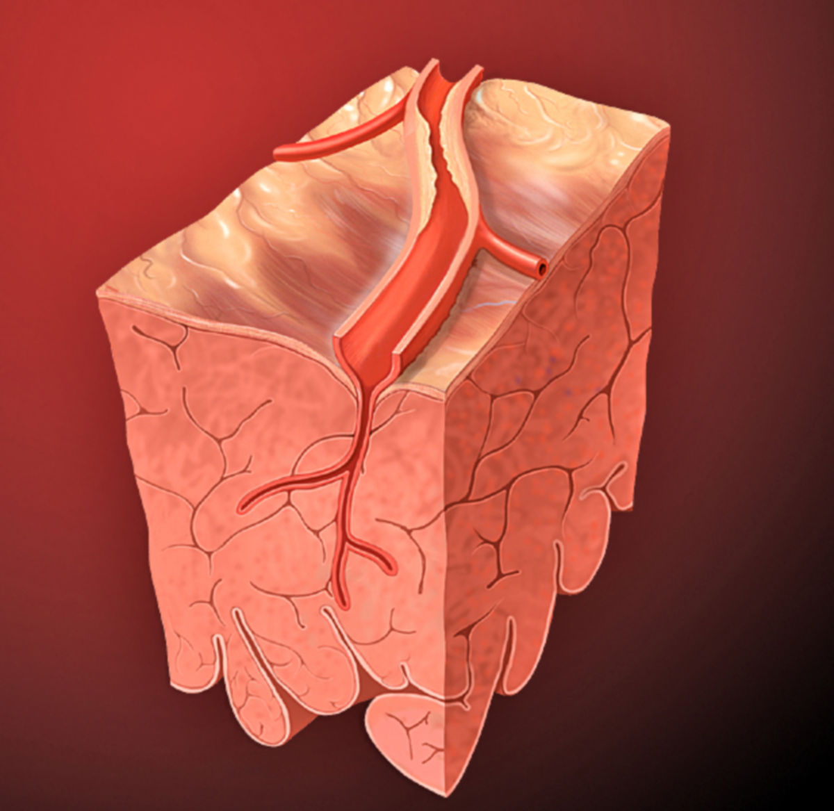
The | The heart’s blood supply comes from two main coronary arteries: the [[Corpus:Left coronary artery|left coronary artery]] (LCA) and the [[Corpus:Right coronary artery|right coronary artery]] (RCA), along with their many branches. | ||
These coronary arteries arise from the ascending part of the [[Corpus:Aorta|aorta]], specifically from the right and left aortic sinuses. | |||
===Branches | === Overview of Coronary Artery Branches: === | ||
* | * '''Left Coronary Artery (LCA, LMCA):''' | ||
** | ** Circumflex branch (RCX, LCx) | ||
** | *** Atrioventricular branches | ||
** | *** Left marginal branch (left marginal artery) | ||
** | *** Left atrial branch (intermediate artery) | ||
** | *** Left atrial anastomotic branch (globular artery) | ||
** | *** Posterior branch of the left ventricle (posterolateral branch) | ||
** | ** Anterior interventricular branch (LAD, left anterior descending artery) | ||
** | *** Conus branch | ||
** | *** Lateral branch (diagonal branch, anterolateral branch) | ||
*** Septal branches (anterior septal branches) | |||
* '''Right Coronary Artery (RCA):''' | |||
** Conus branch | |||
** Sinoatrial node branch | |||
** Atrioventricular branches | |||
** Atrial branches | |||
** Right marginal branch | |||
** Posterolateral branch of the right ventricle (RPLD) | |||
** Atrioventricular node branch | |||
** Posterior interventricular branch (RPD, right posterior descending artery) | |||
** Septal branches (posterior septal branches) | |||
This is just one way to classify the coronary artery branches, as individual anatomy often varies depending on the specific type of blood supply to the heart. | |||
===Arteria coronaria sinistra=== | ===Arteria coronaria sinistra=== | ||
''Abbreviations: LCA, LMCA, ACS'' | ''Abbreviations: LCA, LMCA, ACS'' | ||
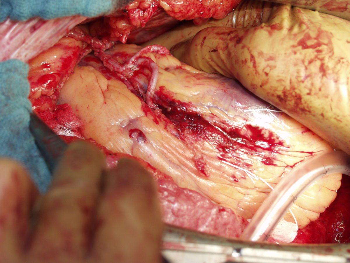
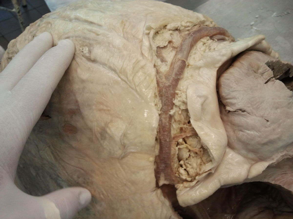
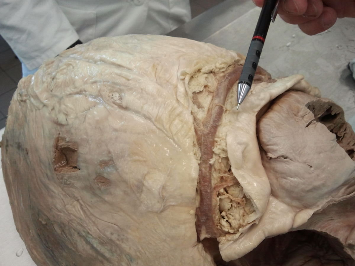
The left coronary artery arises from the left aortic sinus, directly behind the attachment of the aortic valve (valvula semilunaris sinistra). In approximately 75 % of cases, it is more pronounced than the right coronary artery. It runs for a short distance between the conus arteriosus and the left aorta and then splits into two main branches, RIVA and RCX. | The left coronary artery arises from the left aortic sinus, directly behind the attachment of the aortic valve (valvula semilunaris sinistra). In approximately 75 % of cases, it is more pronounced than the right coronary artery. It runs for a short distance between the conus arteriosus and the left aorta and then splits into two main branches, RIVA and RCX. | ||
Version vom 16. Oktober 2024, 16:09 Uhr
This text has been translated by an AI and may sound raw. It will be reviewed shortly. Thank you for your patience!
This text has been translated by an AI and may sound raw. It will be reviewed shortly. Thank you for your patience!
from Latin: corona - wreath, crown
Definition
Anatomy
The heart’s blood supply comes from two main coronary arteries: the left coronary artery (LCA) and the right coronary artery (RCA), along with their many branches.
These coronary arteries arise from the ascending part of the aorta, specifically from the right and left aortic sinuses.
Overview of Coronary Artery Branches:
- Left Coronary Artery (LCA, LMCA):
- Circumflex branch (RCX, LCx)
- Atrioventricular branches
- Left marginal branch (left marginal artery)
- Left atrial branch (intermediate artery)
- Left atrial anastomotic branch (globular artery)
- Posterior branch of the left ventricle (posterolateral branch)
- Anterior interventricular branch (LAD, left anterior descending artery)
- Conus branch
- Lateral branch (diagonal branch, anterolateral branch)
- Septal branches (anterior septal branches)
- Circumflex branch (RCX, LCx)
- Right Coronary Artery (RCA):
- Conus branch
- Sinoatrial node branch
- Atrioventricular branches
- Atrial branches
- Right marginal branch
- Posterolateral branch of the right ventricle (RPLD)
- Atrioventricular node branch
- Posterior interventricular branch (RPD, right posterior descending artery)
- Septal branches (posterior septal branches)
This is just one way to classify the coronary artery branches, as individual anatomy often varies depending on the specific type of blood supply to the heart.
Arteria coronaria sinistra
Abbreviations: LCA, LMCA, ACS
The left coronary artery arises from the left aortic sinus, directly behind the attachment of the aortic valve (valvula semilunaris sinistra). In approximately 75 % of cases, it is more pronounced than the right coronary artery. It runs for a short distance between the conus arteriosus and the left aorta and then splits into two main branches, RIVA and RCX.
In the so-called Bland-White-Garland syndrome, the LCA does not originate in the sinus aortae sinister, but from the left pulmonary artery.
Ramus interventricularis anterior
The anterior interventricular ramus (RIVA, LAD) runs caudally in the anterior interventricular sulcus, runs around the apex of the heart (incisura apicis cordis) and anastomoses in the posterior interventricular sulcus with the posterior interventricular ramus of the right coronary artery. It gives off the following branches:
- Ramus coni arteriosi (RCO): to the conus arteriosus
- Ramus lateralis (ramus anterolateralis, ramus diagonalis, RD): to the anterior wall of the left ventricle (many anatomical variations)
- Rami interventriculares septales (Rami septales anteriores, RSA): extend anteriorly into the interventricular septum and supply it via anterior superior, anterior inferior and apical septal branches. There is also a middle main branch area, from which an artery reaches the anterior papillary muscle of the right ventricle via the moderator ligament and supplies it
Ramus circumflexus
The circumflex ramus (RCX, LCx) follows the left side of the coronary sulcus dorsally to the diaphragmatic facies. Its branches are:
- Ramus nodi sinuatrialis: runs on the anterior side of the left atrium to the right atrium and sinus node
- only fully developed in one third of cases
- can also branch off from the main branch of the coronary sinus artery
- is present in two thirds of cases as ramus atrialis (anterior), which supplies the front of the left atrium and anastomoses with the ramus nodi sinuatrialis of the right coronary artery
- Rami atrioventriculares: branch into rami atriales and rami ventriculares sinistri
- Ramus marginalis sinister: runs downwards at the margo obtusus of the left ventricle
- Ramus atrialis intermedius (ramus atrialis sinister): to the back of the left atrium
- Ramus atrialis anastomoticus (globular artery): small branch that anastomoses with branches of the RCA at the atrial level, can also supply the AV node in some cases
- Ramus posterior ventriculi sinistri (ramus posterolateralis sinister): supplies the facies diaphragmatica of the left ventricle
Versorgungsgebiet
The supply area of the left coronary artery includes:
- anterior wall of the left ventricle (RIVA, lateral ramus)
- partially the anterior wall of the right ventricle (ramus coni arteriosi, RIVA)
- anterior two thirds of the ventricular septum (interventricular septal rami)
- left atrium (rami atriales)
- left ventricle: Lateral wall (ramus marginalis sinister), posterior wall (ramus posterior ventriculi sinistri), with the exception of the area surrounding the posterior interventricular sulcus.
Arteria coronaria dextra
Abbreviations: RCA, ACD
The right coronary artery arises from the ascending branch of the aorta, more precisely from the sinus aortae dexter, directly behind the aortic valve. It runs between the conus arteriosus and the right auricle of the heart to the right section of the coronary sulcus. It follows this and then reaches the facies diaphragmatica of the dorsal heart surface. There it gives off its final branch, the posterior interventricular ramus (RIVP, RPD, R-PDA) at the crux cordis (CC), which runs in the posterior interventricular sulcus towards the apex of the heart and anastomoses with the RIVA of the LCA.
Rami
The RCA gives off the following branches:
- Ramus coni arteriosi: supplies the conus arteriosus and parts of the anterior wall of the right ventricle
- Ramus nodi sinuatrialis: runs on the anteromedial side of the right atrium and usually reaches the sinus node via the sulcus terminalis
- Rami atrioventriculares: atrial and ventricular branches; supply anterior sections of the atrium and ventricle
- Rami atriales: supply lateral sections of the right atrium
- Ramus marginalis dexter: runs along the margo acutus of the right ventricle
- Ramus posterolateralis dexter: runs parallel to the ramus marginalis dexter on the back of the right ventricle
- Ramus nodi atrioventricularis: is released shortly before the exit of the ramus interventricularis posterior, runs in the epicardial fatty tissue under the base of the atrial septum forwards to the AV node
- Ramus interventricularis posterior: terminal branch of the RCA, anastomoses with the RIVA of the LCA
- Rami interventriculares septales (Rami septales posteriores): supply the ventricular septum incl. His bundle and sections of the ventricular conduction system
- anterior superior septal branches: run directly at the exit of the RCA into the upper septum
- posterior superior and inferior septal branches: branch off from the posterior interventricular ramus
Versorgungsgebiet
The supply area of the right coronary artery includes the following structures:
- Sinus node (ramus nodi sinuatralis)
- AV node (ramus nodi atrioventricularis)
- Right atrium (rami atriales)
- Parts of the posterior wall of the left ventricle (ramus posterolateralis dexter)
- Right ventricle: Anterior wall (rami coni arteriosi, ramus marginalis dexter), lateral wall (ramus marginalis dexter), posterior wall (ramus interventricularis posterior)
- Ventricular septum (interventricular septal rami): smaller posterior part, in which the conduction system (e.g. His bundle) is usually localised
Abbreviations
In the clinical context, the full Latin names are rarely used, but mostly abbreviations. English or Latin abbreviations are commonly used.
| English' - short | Latin - short | Long names' |
|---|---|---|
| RCA | ACD | r'ight c'oronary a'rtery A'rteria c'oronaria d'extra |
| LCA, LMCA | ACS | left coronary artery left m'ain coronary artery Arteria coronaria sinistra |
| LAD | RIVA | left anterior descending coronary artery Ramus interventricularis anterior |
| LCx | RCX | l'eft circumflex' coronary artery R'amus circumflex'us |
| RPD, R-PDA | RIVP | right p'osterior d'escending coronary artery R'amus i'nter'v'entricularis p'osterior |
| RPLA | RPLD | right posterolateral artery Ramus posterolateralis dexter |
Supply types
The exact supply areas of the different coronary arteries and their respective branches are subject to significant variations in some cases. For example, the posterior interventricular ramus can originate from the LCA in 20 % of cases and from both coronary arteries in 10 % (codominant supply type).
In approximately 55 % of cases, an intermediate type (normal supply type) is present: In this case, the posterior wall of the heart is supplied approximately equally from the right and left coronary artery.
In 15 to 20 % of cases, the LCA is even more strongly developed (left supply type): The ramus circumflexus then ends as ramus interventricularis posterior on the posterior wall and supplies parts of the right ventricle in addition to the posterior parts of the interventricular septum.
In the right supply type (15 to 25 % of cases), the right coronary artery and in particular the posterolateral dexter ramus are more strongly developed. The latter then supplies most of the posterior wall of the heart and the ventricular septum.
Anatomical variants
Trifurcation
Sometimes an additional artery arises at the bifurcation of the left main artery, between the LAD and LCx, creating a trifurcation. This additional branch is called the ramus intermedius. It runs across the free wall of the left ventricle to the apex of the heart and occurs in about 20% of the population.
Single coronary artery
A single coronary artery (SCA) is a rare congenital anomaly. The Lipton classification categorises the various subtypes of this anomaly into three groups:[1][2].
| group | Course | Name |
|---|---|---|
| I | Follows either the path of a normal right (R) or left (L) coronary artery | RI / LI |
| IIA | Anterior of the pulmonary trunk | RIIA / LIIA |
| IIB | Between the aorta and the pulmonary trunk | RIIB / LIIB |
| IIP | Posterior to the aorta | RIIP / LIIP |
| IIS | Septal course | RIIS / LIIS |
| III | Missing left coronary artery, LAD and RCx arise from the main trunk, starting from the right aortic sinus | RIII |
Physiology
As the heart is dependent on a continuous supply of oxygen due to its high performance, the functionality of the coronary arteries is crucial for the function of the heart muscle. They therefore form numerous anastomoses with each other. However, these are not sufficient to form a complete collateral circulation (functional end arteries). If a branch is blocked, this leads to ischaemia of the supply area with subsequent necrosis of the heart muscle tissue. With increased cardiac work, it is predominantly metabolites (e.g. CO) of the endothelium that mediate vasodilation of the coronary vessels.
Clinic
Diseases
The most important disease of the coronary arteries is arteriosclerosis. It leads to coronary stenosis or gradual occlusion of the coronary arteries - a clinical picture known as coronary heart disease (CHD). Clinically, it manifests itself as angina pectoris, heart attack or heart failure. A distinction is made according to the number of coronary vessels affected:
- Single vessel disease,
- Two-vessel disease,
- three-vessel diseases and
- main stem stenoses
Examination methods
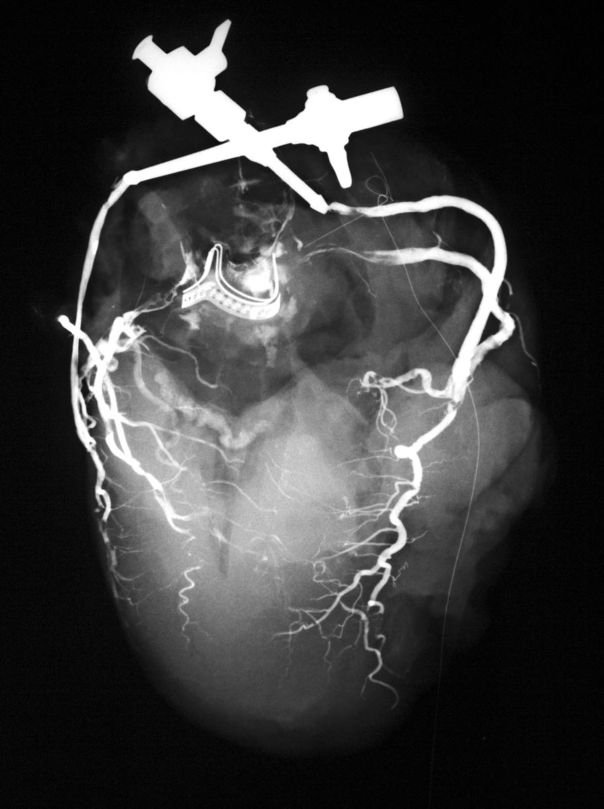
The condition of the coronary arteries is usually checked directly by imaging. The exact extent of the narrowing of the coronary arteries can be determined radiographically using coronary angiography as part of a cardiac catheterisation. Indirect indications of coronary perfusion are provided by examination procedures such as ECG or echocardiography. Further diagnostic procedures are
- Cardio-CT
- Cardio-MRI
- Myocardial scintigraphy
- Intravascular ultrasound (IVUS)
- Coronary angioscopy
Literature
- Köllner, V. / Berg, G.: Behavioural medicine in the prevention and rehabilitation of cardiovascular diseases. In:Herzmedizin 26 (2009). H.2, PP.69-75.
- Herrmann-Lingen, C.: Depression and coronary heart disease. In: Heart Medicine 26 (2009). H.2, P.76-81.
- Benninghoff, Drenckhahn: Anatomy Volume 2, 16th edition 2004, Elsevier: Urban & Fischer
Sources
- ↑ Lipton et al, Isolated Single Coronary Artery: Diagnosis, Angiographic Classification, and Clinical Significance. Radiology, 1979.
- ↑ Katekaru-Tokeshi et al, Applicability of the Leiden Convention and the Lipton Classification in Patients with a Single Coronary Artery in the Setting of Congenital Heart Disease. Journal of Cardiovascular Development and Disease, 2021