Corpus: Transversus thoracis muscle


1. Definition
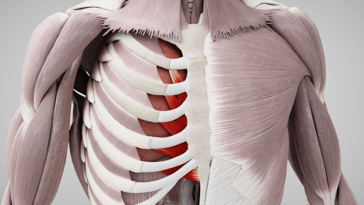
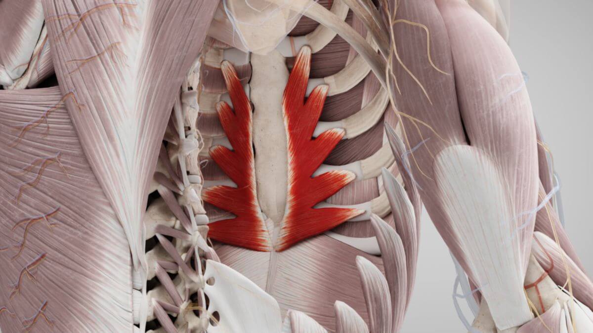
The transversus thoracis muscle is a thin, flat skeletal muscle located on the inner surface of the ribcage, forming part of the intercostal musculature.
2. Progression
2.1. Origin
The transversus thoracis muscle originates from the dorsal surface of the sternum, the back of the xiphoid process, and the sternal parts of the costal cartilages of the 4th to 7th ribs.
2.2. Attachment
The muscle fibers of the transversus thoracis run cranially and laterally, attaching to the lower edge and dorsal surface of the costal cartilages of ribs 2 to 6. The lower (caudal) muscle fibers run almost horizontally and are contiguous with the transversus abdominis muscle, while the upper (cranial) fibers run nearly vertically.
3. Innervation
The transversus thoracis muscle is innervated by the intercostal nerves 2 to 6, corresponding to spinal segments Th2 to Th6.
4. Function
The transversus thoracis muscle maintains dynamic tension in the costal cartilage, which increases the elastic resistance of the thorax. When the muscle contracts, it pulls the costal cartilages downward, thereby assisting in exhalation as an expiratory auxiliary respiratory muscle.
5. Clinic
In cardiac surgery, particularly during coronary artery bypass procedures, the attachment of the transversus thoracis muscle to the upper ribs serves as an important anatomical guide due to its proximity to the internal thoracic artery. This artery is often used in these surgeries.