Corpus: Purkinje fibers


after the Bohemian pathologist and physiologist Jan Evangelista Purkyně (1787–1869)
1. Definition
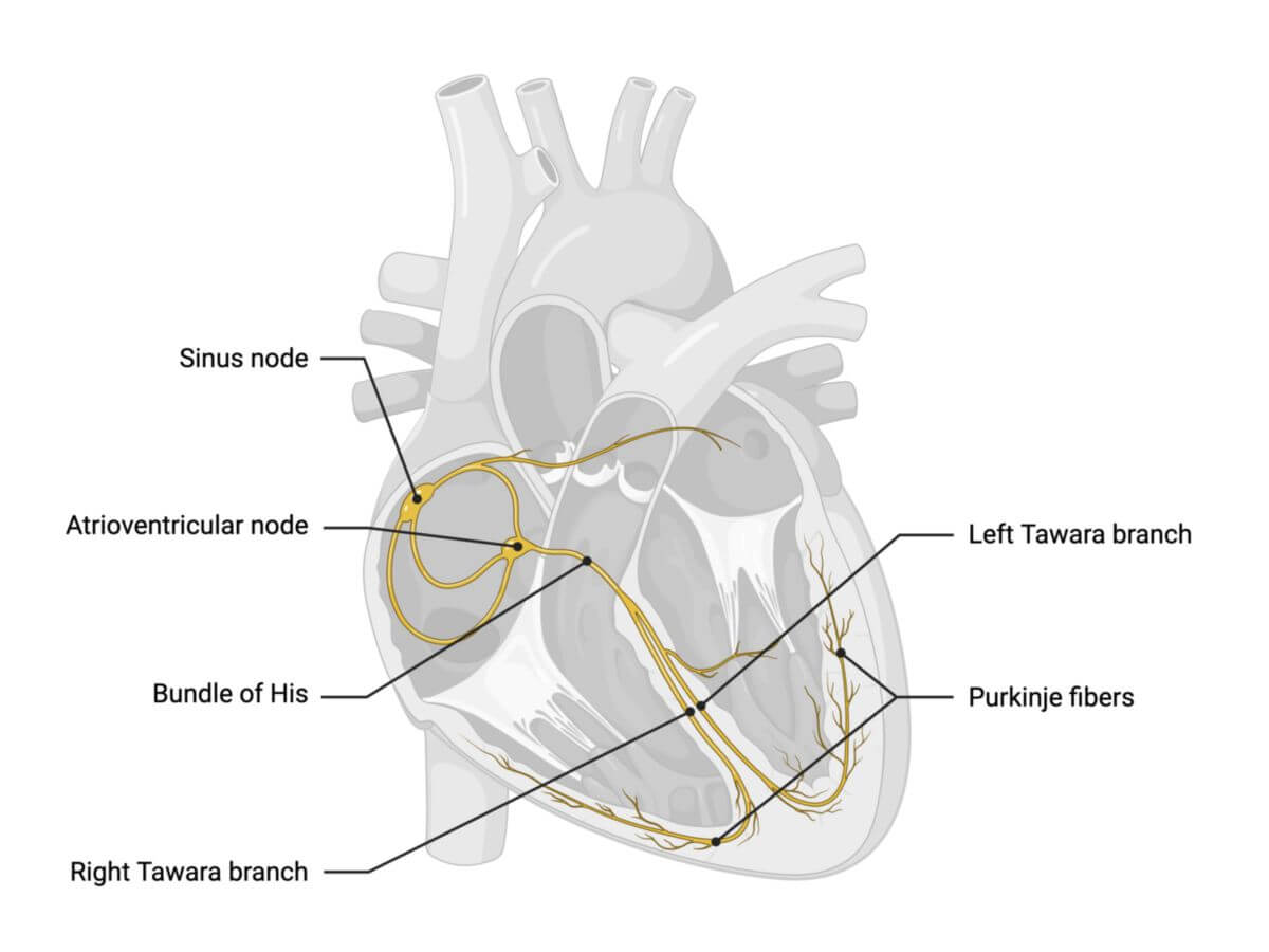
The Purkinje fibres are specialised cardiac muscle cells that represent the final part of the heart's conduction system.
2. Background
Purkinje fibres form the histological foundation of the Tawara branches. Some authors, however, limit the term "Purkinje fibres" specifically to the subendocardial terminal branches of the Tawara branches.
3. Histology
Purkinje fibres are located within the subendocardial tissue. These cells are larger than typical cardiac muscle cells and can be distinguished histologically by their pale cytoplasm. They are rich in glycogen and myofibrils, which can be visualised using Best's carmine staining. Purkinje fibres have fewer mitochondria and T-tubules, which are mainly concentrated at the cell periphery, in contrast to regular heart muscle cells.
4. Physiology
Purkinje fibres are composed of specialised cardiomyocytes that transmit action potentials through gap junctions, primarily using connexins 40 and 43. Their conduction velocity ranges from 2 to 4 m/s, allowing rapid transmission of electrical signals to the ventricular myocardium.
Additionally, Purkinje fibres have the ability to spontaneously depolarise at a lower frequency compared to upstream components of the cardiac conduction system, serving as a final backup pacemaker before activating the working myocardium.
5. Clinic
The time it takes for purkinje fibres to fully excite the ventricular myocardium correlates with the duration of the QRS complex seen on an electrocardiogram.