Corpus: Tawara branch


after the Japanese pathologist Sunao Tawara (1873-1952)
1. Definition
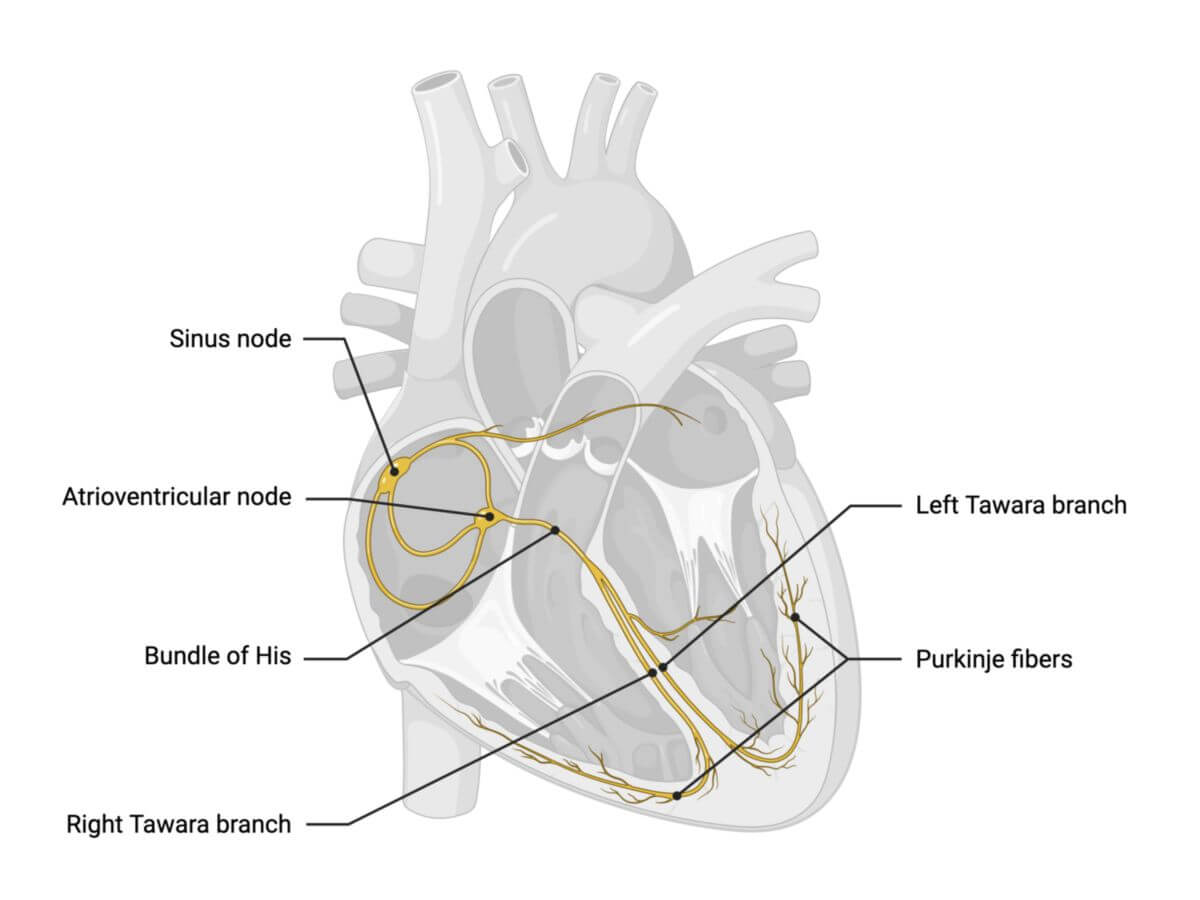
The Tawara branches are part of the cardiac conduction system responsible for transmitting electrical impulses from the His bundle to the working myocardium.
2. Anatomy
The Tawara branch consist of the left and right branches, which emerge from the His bundle and travel through the ventricles.
The left Tawara branch almost vertically and fan-shaped from the His bundle and divides into three fascicles:
- Posterior fascicle: Supplies the posterior papillary muscle and neighboring ventricular myocardium
- Middle fascicle: Extends to the tip of the heart and lateral wall
- Anterior fascicle: Reaches the anterior papillary muscle, apical ventricular septum, and the apex of the heart.
The right Tawara branch runs forward and enters the myocardium of the ventricular septum. It travels subendocardially in the middle third of the septum and radiates to the anterior papillary muscle via the septomarginal trabecula and neighboring ventricular wall. Further branches extend toward the apex of the heart.
.
3. Histology
The Tawara branches consist of specialized cardiomyocytes known as Purkinje fibres. These cells are large, rich in glycogen, and contain myofibrils, though they are only weakly contractile. Purkinje fibres are connected via gap junctions (connexins 40 and 43), while t-tubules are usually absent. Their energy production mainly relies on anaerobic glycolysis.
4. Physiology
The conduction speed within the Tawara legs is 2 to 4 m/s, allowing for the excitation of the entire ventricular musculature within approximately 120 milliseconds. This timing corresponds to the duration of the QRS complex in an ECG.