Corpus: Pia mater


from Greek: pia - soft
1. Definition
The pia mater, part of the leptomeninges, is a connective tissue layer directly covering the brain and spinal cord.
2. Anatomy
The pia mater consists of delicate, soft connective tissue and contains numerous blood vessels. Together with the arachnoid mater, it forms the leptomeninges. The subarachnoid space, filled with cerebrospinal fluid (CSF), is situated between the pia and arachnoid mater.
A topographical distinction is made between:
- Cranial pia mater: surrounds the brain
- Spinal pia mater: surrounds the spinal cord
2.1. Cranial pia mater
The cranial pia mater is a transparent, thin layer of connective tissue with vessels running through it, lying directly on the brain's sulci and gyri. It consists of two layers:
- Inner layer of the pia mater
- Outer layer of the pia mater
The inner layer of the pia mater is located on the superficial glial limiting membrane, formed by astrocyte processes and covered by a patchy basal lamina. This layer consists of loosely interwoven collagen fibril bundles with scattered fibroblast-like meningeal cells.
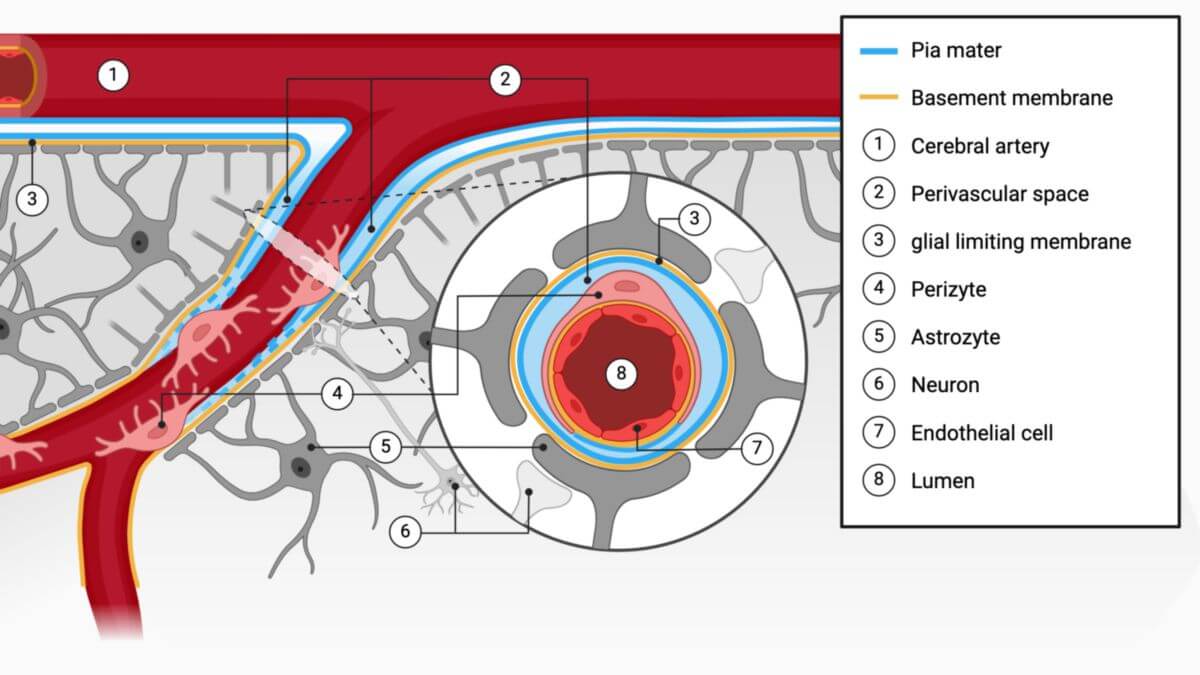
The outer layer of the pia mater is a wide-meshed system of collagen fibril bundles of varying thickness, containing meningeal cells, scattered macrophages, and lymphocytes. It connects to or replaces the adventitia of larger cerebral vessels, following the vessels into the brain substance as far as the pre- and post-capillary sections. This intracerebral, perivascular, adventitial pia space is known as the Virchow-Robin space (perivascular space).
The pia is separated from the subarachnoid space by a mesothelial-like continuous cell layer (leptomeningeal mesothelium), lying on a patchy basal lamina. Mesothelial cells have weakly developed tight junctions, zonulae adherentes, and nexus. This mesothelium does not represent a significant diffusion barrier and extends over the arachnoid trabeculae onto the arachnoid, lining the entire subarachnoid space.
A specialized section of the pia mater is the tela choroidea, forming the lamina propria of the choroid plexus in the cerebral ventricles. In the area of the taenia choroidea, the basement membrane of the plexus epithelium continues into the pia mater’s basement membrane, attaching the plexus to the pia mater. Here, the plexus epithelium merges directly into the ependyma of the ventricles.
The outer layer of the pia mater has its own capillary network, draining via the superficial cerebral veins. Endothelial cells in this area have properties similar to those of the blood-brain barrier. Lymphatic vessels are absent in the pia mater. Sensory, sympathetic, and parasympathetic nerve fibers reach the pia via perivascular plexuses, ending abruptly where the blood vessels enter the brain tissue.
2.2. Spinal pia mater
The spinal pia mater is subdivided into an inner layer (interna) and an outer layer (epipia).
The outer layer contains collagen and elastic fibers, surrounding the spinal cord like a scissor lattice, with reinforcing tracts in the area of the denticulate ligament, the median anterior fissure, and the root fibers’ exit points. These fibers are accompanied by the pia all the way into the dural funnels.
The filum terminale is also covered by the pia, merging with the dura mater in the caudal section and attaching to the periosteum of the second coccygeal vertebra.
The spinal pia mater has its own capillary system, fed by radiculopial arteries from various upstream spinal rami arteries, with anastomoses to the radiculomedullary arteries. Lymphatic vessels are absent. Innervation is provided by the segmental meningeal branches of the spinal nerves.
3. Clinic
Inflammation of the meninges, known as meningitis, can be bacterial or viral. Symptoms include severe fever, vomiting, apathy, and neck stiffness.