Corpus: Choroid plexus


from ancient Greek: χόριον ("khórion") - afterbirth
1. Definition
The choroid plexuses are tangle-like arteriovenous vascular convolutions in the cerebral ventricles composed of specialized glial cells. They are responsible for the production of cerebrospinal fluid (CSF), the formation of the blood-cerebrospinal fluid barrier, and the resorption and detoxification of cerebrospinal fluid.
2. Embryology
The choroid plexus consists of an invagination of the pia mater, covered with ependymal cells and containing blood vessels. The ependymal cells originate from the neural epithelium, which first forms neuroblasts, then glioblasts, and finally ependymal cells. The ependyma lines the neural tube and the later central canal from the inside.
During brain development, the telencephalon grows significantly faster than the diencephalon. Consequently, the soft meninges of both parts of the brain overlay each other, constituting a meningeal duplication called the choroid tela. This connective tissue plate is stretched between the hemispheres and the diencephalon. At its lateral edges, the pia mater forms vascular villi for the plexus of the lateral ventricle and medially covers the roof of the third ventricle. In this area, two rows of vascular villi protrude towards the lumen of the third ventricle, constituting the actual plexus.
The plexus of the fourth ventricle is also formed of a duplication of the pia mater, attaching to the lower surface of the cerebellum and the surface of the hindbrain.
3. Histology
A choroid plexus consists of two layers:
- Epithelial Layer: Plexus epithelium (remodeled hemisphere wall)
- Proper Layer: Vascularized connective tissue of the pia mater (choroid tela)
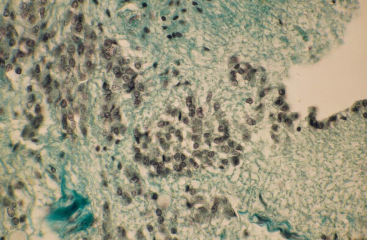
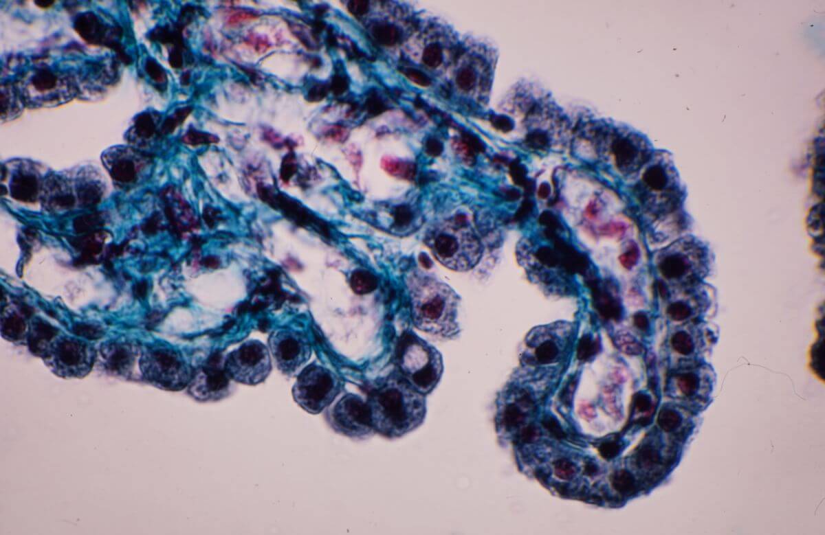
3.1. Plexus epithelium (epithelial layer)
The plexus epithelium consists of specially differentiated glial cells, the ependymal cells. It is cubic and single-layered, but can also be flat or cylindrical in places. At the apical cell poles there are 30 to 60 kinocilia per cell as well as numerous microvilli.
Branched macrophages, so-called epiplexus cells (also known as "Kolmer cells"), adhere to the surface.
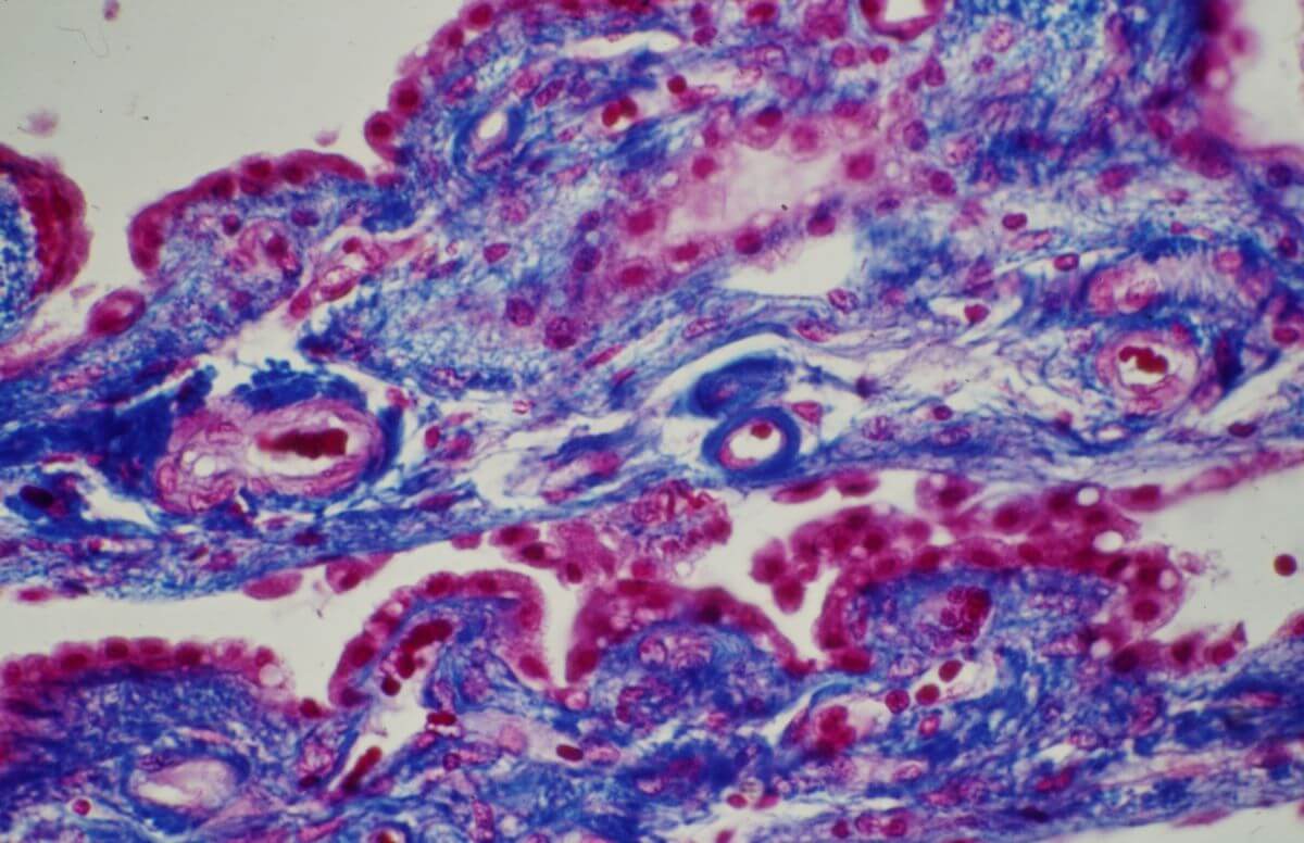
3.2. Choroid Tela (proper layer)
The choroid tela is a specially differentiated form of the pia mater, rich in collagen fibers and heavily interspersed with capillaries. The capillaries have a fenestrated endothelium and a continuous basal lamina that constitutes labyrinthine branches penetrating the surrounding connective tissue. Collagen formations within the connective tissue can calcify to constitute psammoma bodies.
4. Anatomy
The choroid plexus is found:
- On the inside of the inferior horn and the central part of the lateral ventricles
- In the roof of the third and fourth ventricles
At the lateral end of the fourth ventricle, part of the choroid plexus protrudes from the lateral aperture (foramen of Luschka) on both sides, known as Bochdalek's flower basket due to its shape. The choroid plexus of the lateral ventricles is connected to that of the third ventricle via the interventricular foramen (foramen of Monro). Its point of attachment to the thalamus is called the choroid line.
5. Function
The choroid plexus fulfills three main functions:
5.1. Formation of cerebrospinal fluid
The active transport of sodium ions through the sodium-potassium ATPase in the luminal plasma membrane of plexus epithelial cells drives CSF production. Chloride ions and water follow the sodium ions through specific ion channels and various chloride channels. In addition to ions, the plexus secretes glucose, vitamins C and B12, various nucleosides, and other substances, including leptin and transthyretin (a transport protein for thyroid hormones).
5.2. Barrier function
The blood-cerebrospinal fluid barrier includes the fenestrated endothelium, the endothelial basement membrane, and the plexus epithelial cells (with tight junctions) with their own basement membrane. Small hydrophilic molecules and peptides can pass through the fenestrated sections of the endothelium, but larger particles cannot.
5.3. Resorption and detoxification of the cerebrospinal fluid
The plexus epithelial cells contain various transporters and molecular detoxification systems. These include the multi-drug resistance transporter 1 (MDR1), which transports pharmaceuticals (especially barbiturates), bilirubin, bile salts, and leukotrienes into the choroid tela (i.e., into its blood vessels).
Transporters for organic cations and anions, as well as glutamate (e.g., EAA3 transporter), have also been detected.