Corpus: Orbit
Achtung: Du siehst nicht die aktuelle, sondern eine ältere Version dieser Seite.


Synonym: orbital cavity; orbit; eye socket
This text has been translated by an AI and may sound raw. It will be reviewed shortly. Thank you for your patience!
This text has been translated by an AI and may sound raw. It will be reviewed shortly. Thank you for your patience!
1. Definition
The orbit is the cavity located in the frontal region of the skull in which the eye with its appendages and the blood vessels and nerves leading to and from it are located.
2. Anatomy
2.1. Overview
The orbit is formed by parts of various skull bones. It has roughly the shape of a four-sided pyramid, with its base pointing forwards and its tip pointing into the depths of the skull. It forms a bony protective and receptive shell for the eye. The bones involved in the structure of the orbit are listed below:
- zygomatic bone
- frontal bone
- lacrimal bone
- upper jaw
- ethmoid bone
- palatine bone
- sphenoid bone
2.2. Limitations
The orbital roof is formed anteriorly by the Os frontale (Facies orbitalis ossis frontalis) and posteriorly by the Os sphenoidale (Ala minor ossis sphenoidalis).
The orbital floor consists mainly of bone parts of the maxilla (Facies orbitalis corpus maxillae) and the Os zygomaticum. A small area of the floor in the posterior part of the orbit forms the palatine bone (processus orbitalis ossis palatini). The lowest point of the orbital floor is called the orbital bone.
The lateral orbital wall forms the zygomatic bone (facies orbitalis) and the sphenoid bone (ala major ossis spenoidalis).
The very thin medial orbital wall is formed from front to back by the maxilla, the Os lacrimale, the Os ethmoidale (Lamina papyracea), the Facies orbitalis ossis frontalis and the Ala minor ossis sphenoidalis.
2.3. Openings
The frontal access to the orbit, i.e. the orbital opening, is also known as the aditus orbitalis. It is bordered by the bony orbital rim (margo orbitalis).
The orbit is connected to the middle cranial fossa by the superior orbital fissure and the optic canal. The inferior orbital fissure connects it to the pterygopalatine fossa. A large number of important pathways reach the orbit through both fissures (see there).
The Os lacrimale and the Maxilla form - limited by the Crista lacrimalis anterior and posterior - the Fossa sacci lacrimalis and the Canalis nasolacrimalis, which harbours the nasolacrimal duct (Ductus nasolacrimalis).
The infraorbital sulcus forms the entrance to the infraorbital canal for the nerves and vessels of the same name.
The anterior ethmoidal foramen and the posterior ethmoidal foramen allow the nerves and vessels of the same name to return from the orbit into the cranial cavity.
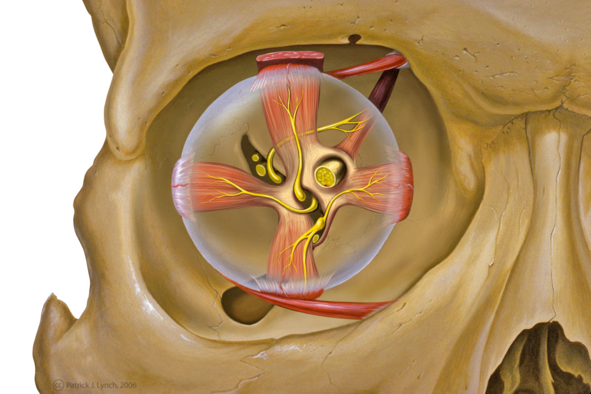
2.4. Contents
The following structures are located in the orbit:
- Bulbus oculi (eyeball)
- Eye muscles
- Retrobulbar fat body
- Lacrimal apparatus: Glandula lacrimalis (lacrimal gland), ductus nasolacrimalis (nasolacrimal duct), saccus lacrimalis (lacrimal sac)
- Nerves: optic nerve (optic nerve), ophthalmic nerve, oculomotor nerve, trochlear nerve, abducens nerve. Nervi ciliares breves
- Blood vessels: ophthalmic artery and vein
- Ganglia: ciliary ganglion
3. Histology
The orbit consists of the typical bony parts of the flat bones of the skull. The bony orbit is separated from the contents by a layer of periosteum, which is referred to here as the periorbita.
4. Function
The orbit serves to hold, fix and protect the eye and as the origin of the eye muscles.
5. Clinic
The orbit is often involved in midface fractures. This can lead to a stepped formation of the orbital rim. The direct impact of force on the eye can rupture the orbital floor. This is referred to as a blow-out fracture.
The orbit and its contents are also affected in a number of autoimmune diseases. Typical examples of these so-called orbitopathies are myositis of the eye muscles and Graves' disease.