Corpus: Optic tract
1. Definition
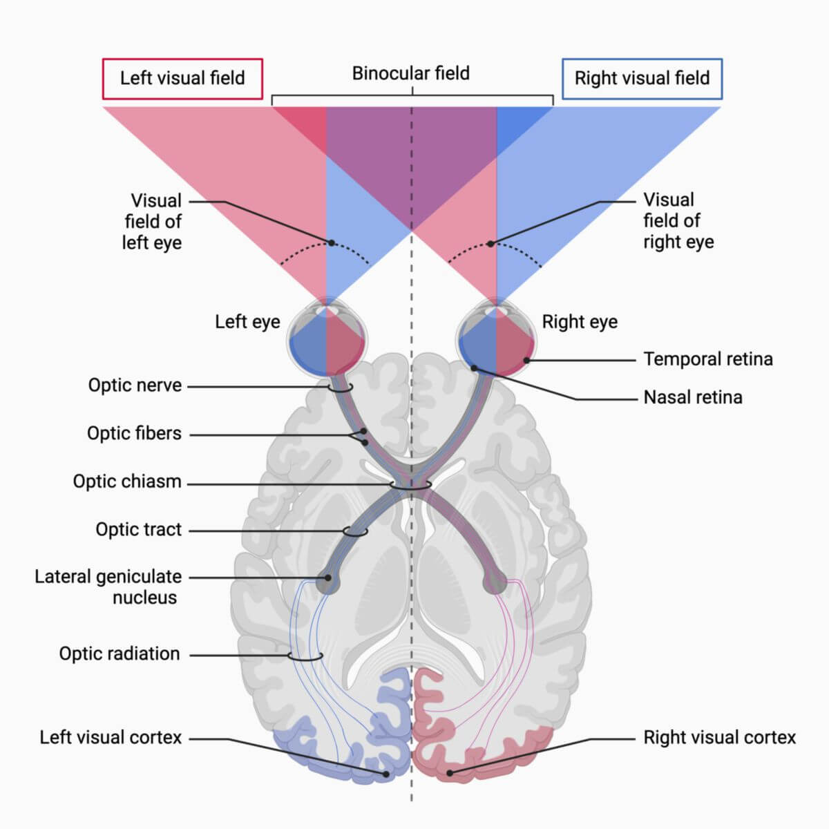
The optic tract is the section of the visual pathway between the optic chiasm and the lateral geniculate nucleus. It contains neurons from the ipsilateral temporal and the contralateral nasal parts of the retina. Accordingly, the transmitted visual impressions originate from the contralateral visual field.
2. Background
Visual signals are transmitted to the visual cortex via several stations in the visual pathway.
Rods and cones of the retina form the first neuron of the visual pathway in the outer granular layer as receptor cells. Transmission continues to the second, bipolar neuron in the inner granular layer. The third, multipolar neuron (ganglion cell) is located in the ganglion cell layer of the retina.
The axons of the ganglion cells constitute the optic nerve and run through the optic canal to the optic chiasm. There, the fibers originating from the nasal part of the retina (i.e., temporal visual field) cross to the opposite side. They then travel with the uncrossed optic nerve fibers of the other eye in the optic tract to the thalamus, specifically to the lateral geniculate nucleus (LGN).
Approximately 1 million axons travel from the LGN as optic radiations to the neurons of the primary visual cortex (striate cortex or V1, Brodmann area 17).
About 10 % of the optic fibers bypass the LGN and travel directly from the optic tract to the hypothalamus, diencephalon (pretectal nuclei), and via the superior brachium to the midbrain (superior colliculi). This serves the accommodation and pupillary reflex, circadian rhythm, and reflex visual motor function (extrastriate visual pathway).


