Corpus: Electroencephalography

1. Definition
Electroencephalography, or EEG for short, is a neurological diagnostic procedure used to record electrical activity in the brain. This activity is displayed graphically as brain waves, which represent the collective electrical signals of nerve cells. The recorded results are called an electroencephalogram.
2. Performance
Electrodes are placed on the scalp to continuously detect, amplify, and record fluctuations in electrical potential (brain waves) in the brain. In neurology, at least 12 leads are typically used.
3. Indications
The primary use of electroencephalography is the diagnosis of epilepsy. The sensitivity of a routine EEG performed between seizures (interictal EEG) is estimated to be around 30–50%, meaning that in many cases, the EEG of epilepsy patients appears normal. Despite this limitation, EEG remains the gold standard for confirming epilepsy.
EEG is also used to assess brain death.
In sleep medicine, EEG is an integral part of polysomnography. During sleep studies, EEG is recorded using a reduced number of electrodes throughout the night. The dominant wave patterns help determine sleep stages, while specific features such as sleep spindles and K-complexes are characteristic of certain sleep phases.
4. Assessment
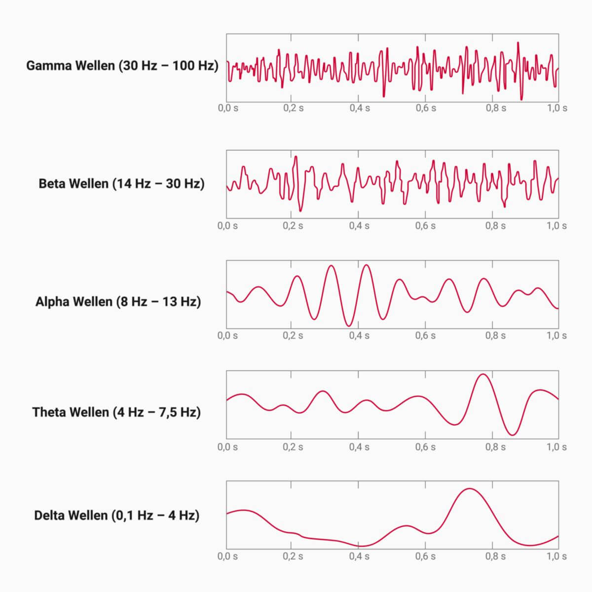
EEG interpretation is based on the frequency of brain waves, which are categorized into specific frequency bands. The exact classification varies across different sources, as it is based on historical categorization, some of which has been revised with modern research.
A positive potential shift, seen as a downward deflection on the EEG, results from excitatory postsynaptic potentials (EPSPs) in the deeper cortical layers (lamina IV) or inhibitory postsynaptic potentials (IPSPs) in the superficial cortical layers. Conversely, a negative potential shift, seen as an upward deflection on the EEG, may result from EPSPs in the dendrites of the superficial cortex or IPSPs in the deeper layers.
5. Pathological EEG findings
- General changes
- Persistent slowing of brain waves
- Diffuse and paroxysmal dysrhythmias
- Seen in conditions such as inflammation and neurodegenerative disorders
- Focal abnormalities
- Localized wave changes
- Seen in stroke, hematomas, and other focal brain lesions
- Epileptiform activity
- Characteristic findings in epilepsy
- Includes spikes, sharp waves, and spike-and-wave complexes
- Flat EEG (Isoelectric line)
- Criterion for brain death
- Absence of detectable electrical activity
6. Other diagnostic procedures
In addition to EEG, magnetoencephalography (MEG) is used to measure the magnetic fields generated by neuronal activity in the brain.