Corpus: Ulnar nerve


Definition
The ulnar nerve is a mixed motor and sensory nerve of the arm that arises from the medial fasciculus of the brachial plexus. It contains fibers from C8 and Th1.
Course
The ulnar nerve runs along the medial side of the arm to the forearm and hand. It originates medial to the axillary artery and accompanies the emerging brachial artery in the medial bicipital groove to approximately the middle of the upper arm. There it penetrates the medial intermuscular brachial septum, passes the medial head of the triceps brachii muscle and travels to the ulnar nerve groove between the medial epicondyle and the olecranon of the ulna.
At the elbow, it continues to attach to the posterior of the medial epicondyle and reaches the forearm between the two muscle heads of the flexor carpi ulnaris muscle. There, the nerve continues distally on the ulnar side of the flexor digitorum profundus muscle. In the upper 1/3 of the forearm, it is still covered by the flexor carpi ulnaris muscle and is separated from the ulnar artery. In the distal 2/3, it runs close to the medial side of this artery.
In front of the wrist, the nerve divides into a superficial and a deep branch. The deep branch joins the ulnar artery at the wrist above the transverse carpal ligament through the so-called ulnar canal. It is laterally restricted by the pisiform bone and palmarly by the palmar carpal ligament and the palmaris brevis muscle.
Branches on the forearm
- articularis cubiti branches
- muscular branches
- palmar branches
- dorsal branches
- dorsal digital nerves
Branches on the hand
- superficial branches (mixed, predominantly sensitive)
- muscular branch to the palmaris brevis muscle
- common palmar digital nerves, from which two proper palmar digital nerves emerge
- deep branch (strictly motor)
Anastomoses
The connection between the ulnar nerve and the median nerve is known as a Martin-Gruber anastomosis. It is a common anatomical variant.
Function
Motor innervation
The ulnar nerve innervates parts of the forearm muscles, supporting flexion of the finger joints and wrist. Its main supply area is the hand muscles. It supplies large parts of the thenar (heel of thumb) and hypothenar muscles (heel of little finger), as well as most of the short muscles of the middle hand, which are responsible for spreading and closing the fingers.
Forearm
Hand
- interossei palmares muscles
- interossei dorsales muscles
- lumbricales muscles (third and fourth)
- abductor digiti minimi muscle
- flexor digiti minimi brevis muscle
- opponens digiti minimi muscle
- adductor pollicis muscle
- flexor pollicis brevis muscle, deep head
- palmaris brevis muscle
Sensory innervation
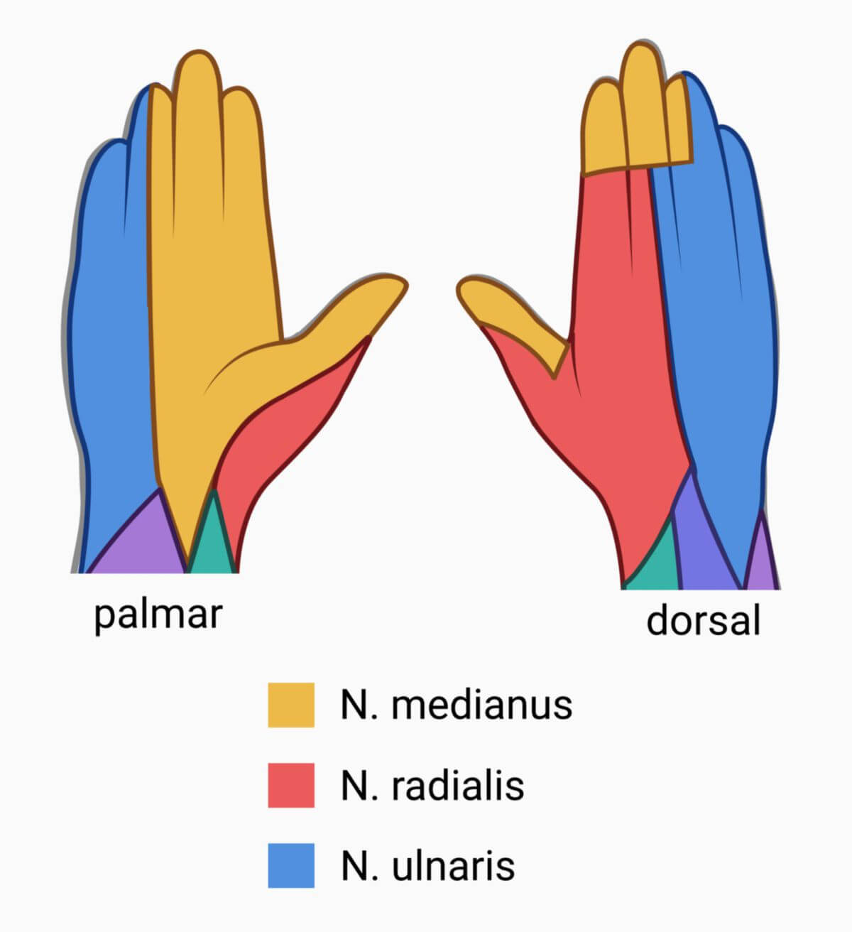
The ulnar nerve sensory supplies the skin above the heel of the little finger and the corresponding region on the ulnar side of the back of the hand. Its terminal branches supply the little finger completely and the ulnar part of the ring finger. Additionally, it supplies the radial part of the ring finger on the dorsal side and the ulnar half of the middle finger to about the middle phalanx.
Clinic
The ulnar nerve can easily be injured by trauma (e.g., fracture) to the elbow. Even a push or slap on the nerve causes an electric shock-like sensation in the arm.
The passage of the nerve through the ulnar nerve groove is a constriction that can be the cause of ulnar groove syndrome.
Complete failure of the ulnar nerve (ulnar nerve palsy) leads to paralysis of the short finger muscles and thus to the clinical picture of the claw hand.
Ulnar tunnel syndrome refers to damage to the ulnar nerve on the ulnar side of the wrist in the area of the so-called Guyon's tunnel.
The ulnar nerve is the most commonly used nerve for relaxometry.