Corpus: Recurrent laryngeal nerve
Achtung: Du siehst nicht die aktuelle, sondern eine ältere Version dieser Seite.


This text has been translated by an AI and may sound raw. It will be reviewed shortly. Thank you for your patience!
This text has been translated by an AI and may sound raw. It will be reviewed shortly. Thank you for your patience!
Synonyms: recurrent nerve, vocal nerve
1. Definition
The recurrent laryngeal nerve is a paired branch of the vagus nerve. The section of the recurrent laryngeal nerve close to the larynx is also referred to as the inferior laryngeal nerve.
2. Core areas
The special visceromotor fibres of the recurrent laryngeal nerve originate from the ambiguous nucleus and emerge from the brain stem with the cranial root of the accessorius nerve. They then cross as the internal ramus to the vagus nerve, which receives them in the jugular foramen. The cell bodies of the sensory nerve fibres are located in the ganglion inferius nervi vagi (ganglion nodosum) - from there they run to the nucleus solitarius. The parasympathetic fibres originate from neurons of the dorsal vagus nerve nucleus.
3. Course
The right and left recurrent laryngeal nerves differ in their course.
The left recurrent laryngeal nerve (sinister) originates in the upper mediastinum and loops around the aortic arch (arcus aortae). It then runs through the oesophageotracheal sulcus between the oesophagus and trachea to the larynx.
The right recurrent laryngeal nerve (dexter) leaves the vagus nerve at the superior thoracic aperture. It loops around the subclavian artery (dextra) and runs cranially to the side of the trachea in the oesophageotracheal sulcus.
The nerves on both sides cross the inferior thyroid artery on their way to the larynx and finally enter the larynx from the dorsal side between the cricoid cartilage and thyroid cartilage. In doing so, they pierce the constrictor pharyngis medius muscle.
4. branches
The recurrent laryngeal nerve gives off the following branches:
- Rami oesophageales, supplying the cervical part of the oesophagus
- Rami tracheales to the cervical section of the trachea
The final branch of the recurrent laryngeal nerve is the inferior laryngeal nerve. It anastomoses with the superior laryngeal nerve in the Galen anastomosis.
Some textbooks list additional inferior cardiac nerves with a connection to the sympathetic trunk, which run to the cardiac plexus.
5. Variety
In rare cases (about 1:200), the right nerve does not form a loop around the right subclavian artery. It then branches off from the vagus nerve at the level of the cartilago cricoidea. This course is frequently associated with a vascular variant in which the right subclavian artery branches off on the left side of the aorta and then changes to the right behind the oesophagus.
6. Embryology
The loop-like course of the recurrent laryngeal nerve is caused by the "descent" of the heart into the thorax during embryonic development. It is the nerve of the 6th gill arch.
7. Function
The recurrent laryngeal nerve innervates almost all laryngeal muscles (with the exception of the cricothyroid muscle), i.e:
- posterior cricoarytenoid muscle
- lateral cricoarytenoid muscle
- arytenoid transversus muscle
- arytenoid oblique muscle
- thyroarytenoid muscle
These muscles open and close the glottis by abducting and adducting the vocal folds. The nerve therefore plays an essential role in voice production (phonation) and breathing.
The sensitive parts of the recurrent laryngeal nerve supply the mucous membrane of the larynx below the glottis (subglottic space).
Parasympathetic fibres from the vagus nerve travel to the tracheal glandulae in the upper section of the trachea and regulate their secretion production.
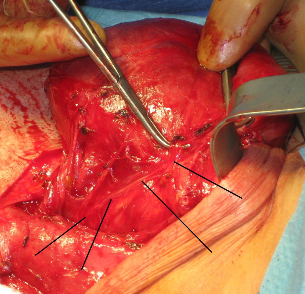
8. Clinic
A unilateral or bilateral loss of the recurrent laryngeal nerve leads to so-called recurrent nerve palsy. It can occur, for example, as a complication of a thyroidectomy or other operations in the throat area. Other causes are
- Tumours in the mediastinum
- Pancoast tumours
- Aortic aneurysm
Unilateral paralysis manifests itself as hoarseness, while bilateral paralysis leads to severe dyspnea.
9. Literature
- Waldeyer et al. Human anatomy: Textbook and Atlas in One Volume (De Gruyter Studium) (19th totaly rev. ed.), De Gruyter, 2012