Corpus: Palatine tonsil


1. Definition
The palatine tonsil is an almond-shaped lymphoid organ localized between the anterior and posterior palatine arches.
2. Anatomy
2.1. Topography
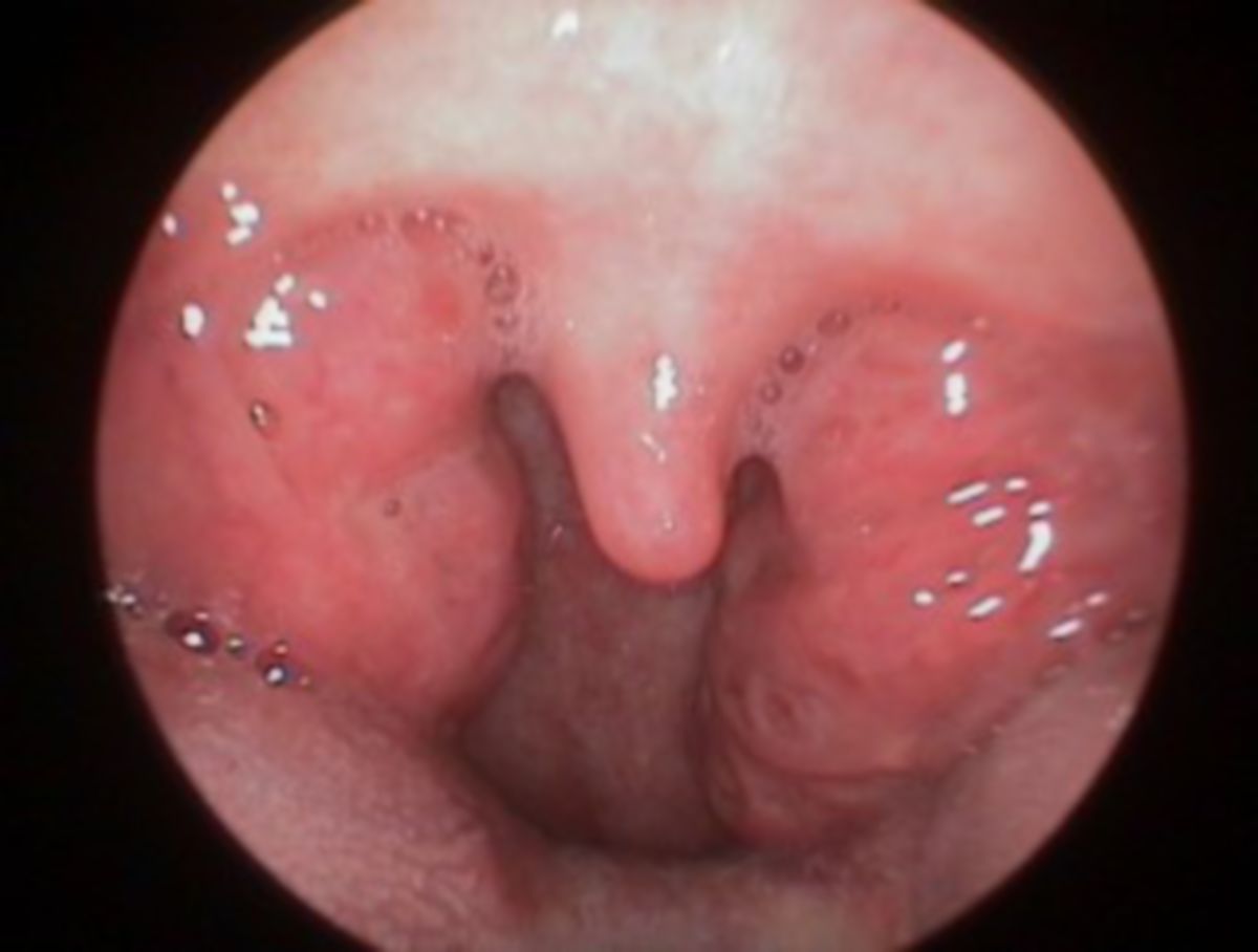
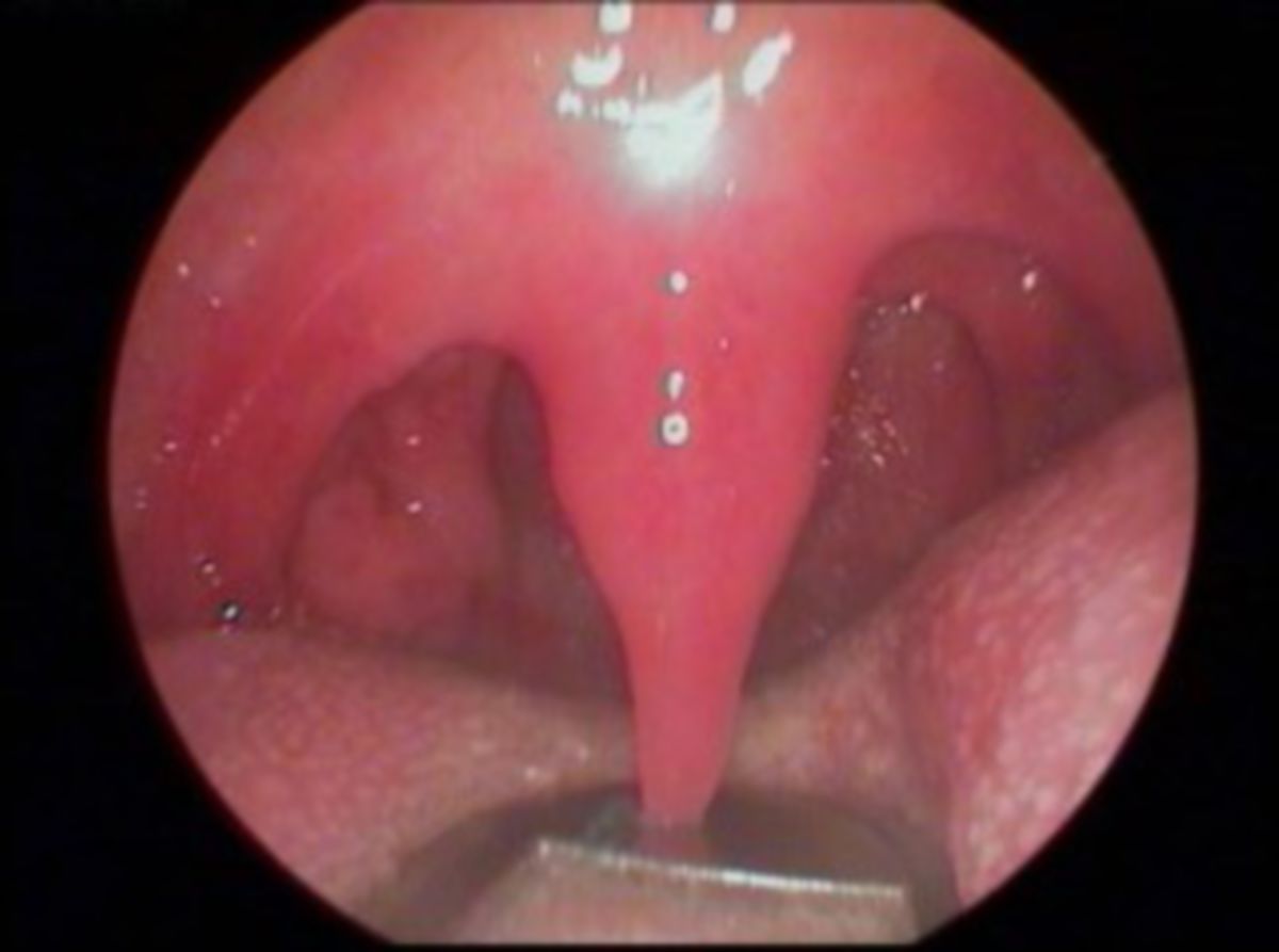
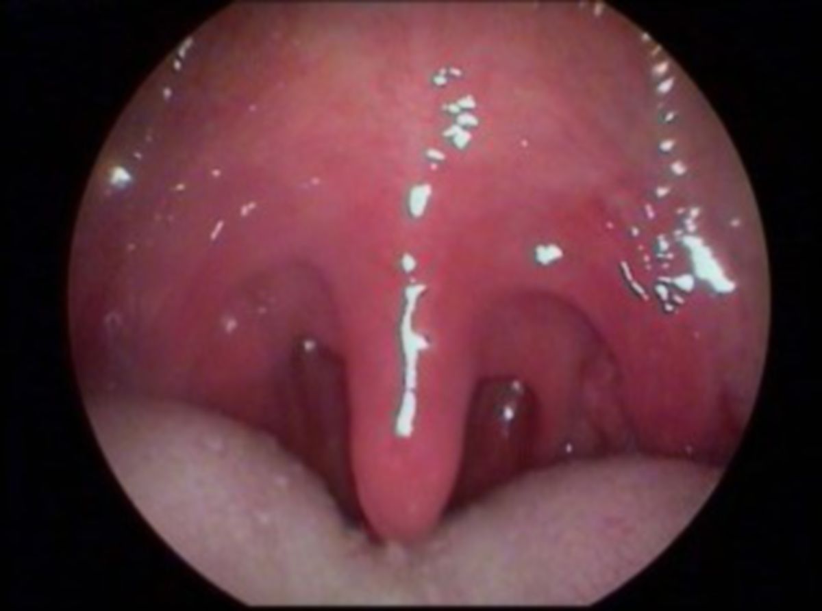
The palatine tonsils, integral to Waldeyer's pharyngeal ring, are bilaterally situated in the oral cavity's posterior region at the pharyngeal isthmus, marking the transition to the pharynx. Positioned between the palatoglossal and palatopharyngeal arches, this area is termed tonsillar fossa, where the tonsils are ensconced within a connective tissue capsule.
Adjacent to the palatine tonsil is the superior constrictor muscle of the pharynx.
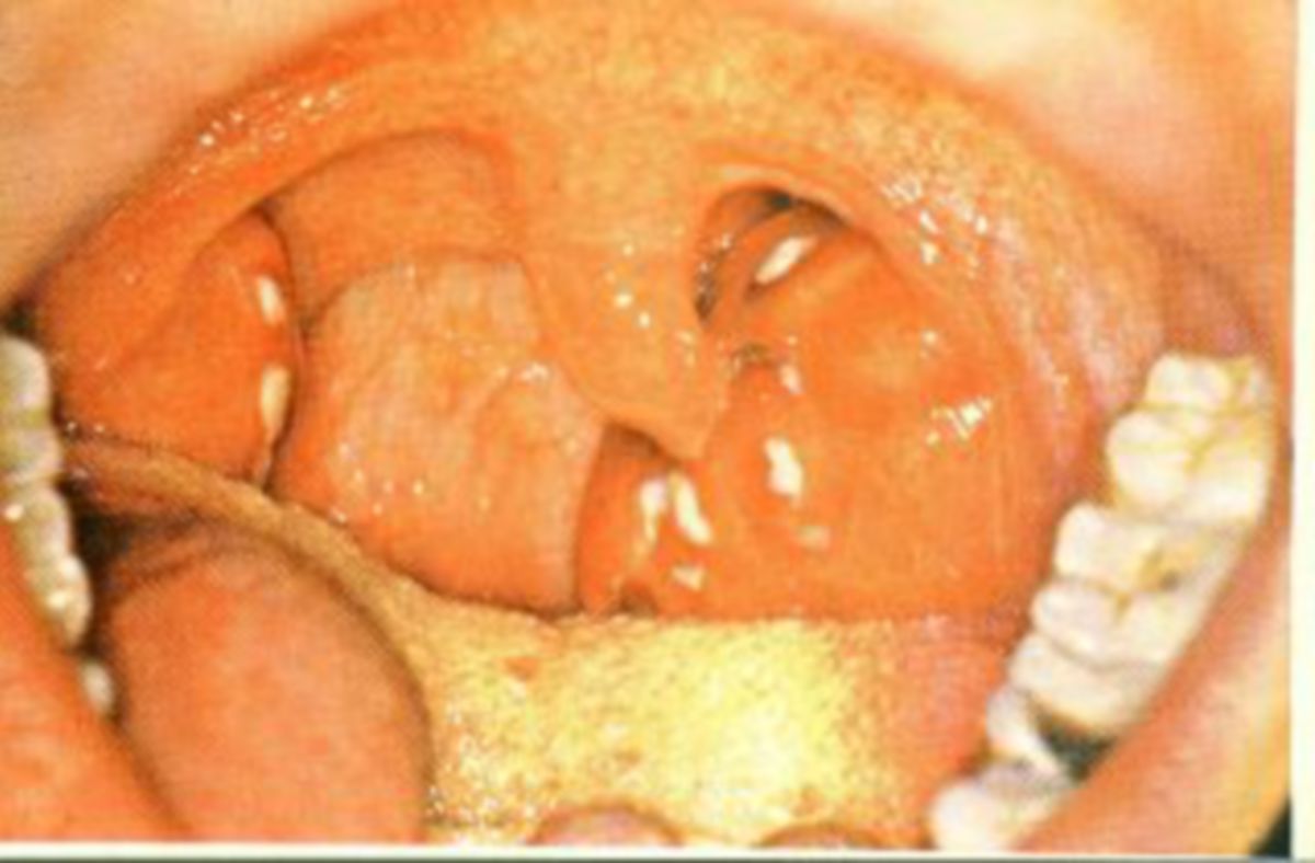
2.2. Structure
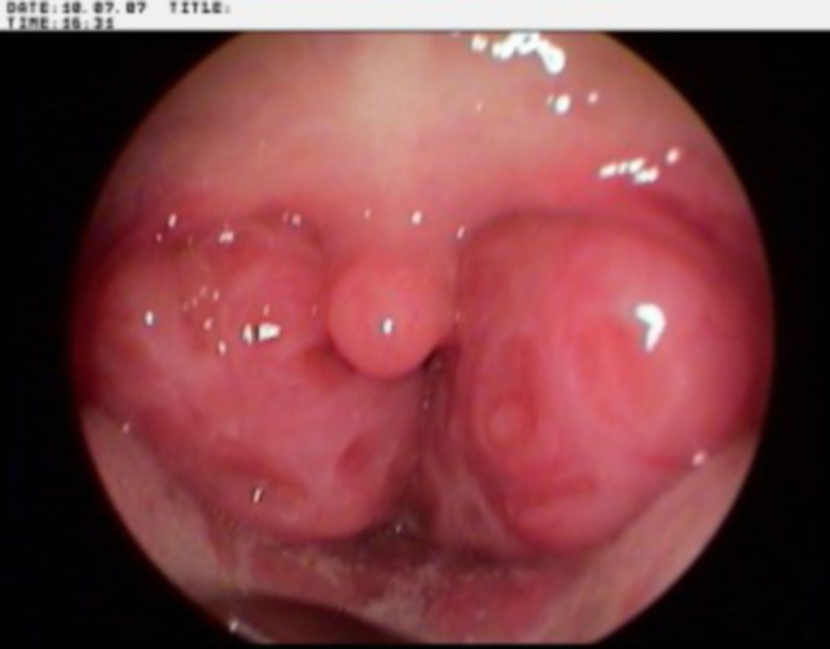
Characteristically ellipsoid, the palatine tonsil measures approximately 2-3 cm in length and 1-2 cm in width in adults. The side facing the pharyngeal isthmus features 10-15 small openings known as tonsillar pits. From these openings, deep crypts (cryptae tonsillae) extent, lined with multi-layered, non-keratinized squamous epithelium that significantly enlarges the surface area to about 300 cm² per tonsil.
Beneath the epithelial layer lies lymphoreticular connective tissue filled with numerous primary and secondary follicles. The surface-facing side of the secondary follicles shows dense clusters of lymphocytes, referred to as lymphocyte caps.
The crypts, closely spaced and branched, impart a serrated appearance to the palatine tonsil. Food particles, along with shed epithelial cells and leukocytes, occasionally accumulate in these crypts, forming the so-called tonsillar plugs or "tonsilloliths," which may appear as white protrusions on the tonsil's surface.
Surrounding the palatine tonsil are mucous salivary glands, typically opening adjacent to the tonsil.
2.3. Vascular Supply
The palatine tonsil's arterial supply varies. It is predominantly provided by the tonsillar branch of the ascending palatine artery, with contributions from direct tonsillar branches of the facial artery and minor palatine arteries from the descending palatine artery. Some parts also receive blood from the dorsal lingual branches of the lingual artery.
Venous drainage occurs through the pharyngeal venous plexus and the internal jugular vein.
Lymphatic drainage is managed by the submandibular lymph nodes and drains into the deep cervical lymph nodes.
2.4. Innervation
Sensitive innervation of the palatine tonsils is primarily via fibers from the glossopharyngeal nerve (IX cranial nerve) and the vagus nerve (X cranial nerve). They also receive sensory fibers from the maxillary nerve (V2) through the lesser palatine nerves, which pass through the pterygopalatine ganglion.
3. Embryology
The tonsil bay and the palatine tonsil originate from the second pharyngeal pouch, an invagination between the second and third pharyngeal arches. Epithelial buds from this pouch's endoderm proliferate into the surrounding mesenchyme, which differentiates into lymphatic tissue by the twentieth week of gestation.
4. Histology
Like its counterparts, the palatine tonsil consists of lymphatic tissue dotted with numerous lymph follicles. Its pharyngeal side is covered by squamous epithelium that extends deep into the crypts. Here, the epithelium is looser, and the basement membrane is partially absent, facilitating robust antigen-immune cell interaction.
The palatine tonsil is encased in a connective tissue capsule, from which septa extend into the interior, further dividing the tonsil into lobules.
5. Immunology
As part of the mucosa-associated lymphoid tissue (MALT), the palatine tonsil plays a vital role in the immune system. Along with the other tonsils, it acts as a defensive barrier against pathogens attempting to enter the throat via the oral mucosa. This function is crucial in preventing potential infections of the gastrointestinal and respiratory tracts.
The B lymphocytes of the palatine tonsil can produce all five classes of immunoglobulins. When exposed to antigens like diphtheria toxin, polioviruses, Streptococcus pneumoniae, Haemophilus influenzae, or Staphylococcus aureus in vitro, they rapidly generate specific antibodies. Additionally, a significant T cell response is elicited, marked by increased cytokine production by T helper cells.
6. Clinic
Tonsillitis, inflammation of the palatine tonsil, ranks among the most frequent ENT disorders, predominantly affecting children and presenting in both acute and chronic forms. With a more cautious approach to tonsillectomy today compared to past decades, due to the tonsils' immunological significance, the procedure is less frequently recommended.
Unphysiological enlargement of the palatine tonsil without inflammation is known as tonsil hyperplasia.