Corpus: Metacarpal bone

1. Definition
The 5 metacarpal bones form the bony basis of the metacarpus.
2. Division
The metacarpal bones are simply numbered systematically from lateral to medial for their correct anatomical designation, with the slightly splayed first metacarpal bone carrying the thumb:
- first metacarpal bone
- second metacarpal bone
- third metacarpal bone
- forth metacarpal bone
- fifth metacarpal bone
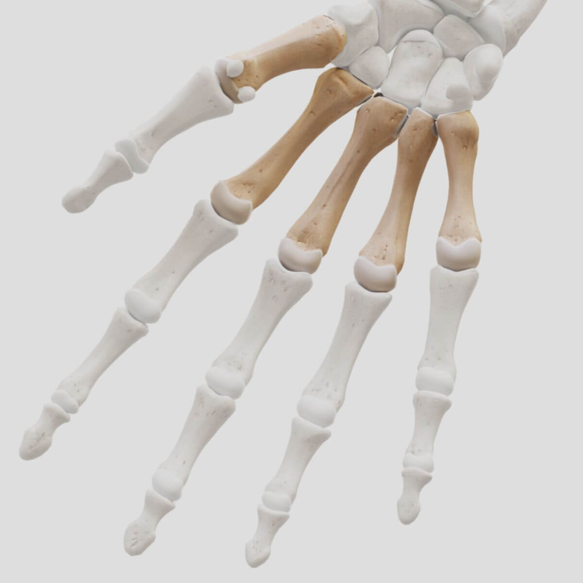
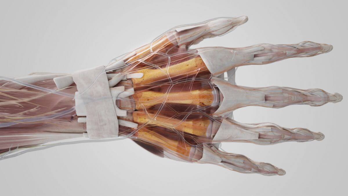
The metacarpal bones II and III are the longest, the metacarpal bone I is the shortest. The spaces between the metacarpal bones are known as Intermetacarpal spaces. They are filled by the interosseous muscles.
3. Anatomy
3.1. Common features
All metacarpal bones consist of three sections. Seen from proximal to distal, these are:
- base
- shaft
- head
The base of the metacarpals has a cubic basic shape. The concave cartilage surfaces of the bases articulate with the carpal bones in the carpometacarpal joints. Between the metacarpal bones II to V, smaller cartilage facets form additional amphiarthroses, the intermetacarpal joints. The metacarpal joint is formed from the two joint types as a secondary joint. The shaft has a triangular cross-section. The tip of the triangle points towards the palm, the base towards the back of the hand. In the distal section of the metacarpal bones, this side is an almost flat bony surface over which the tendons of the extensors run. The other two sides, i.e., the medial and lateral sides of the metacarpal bone, are concave. This is where the interosseous muscles attach. At the front they meet in a bone ridge. The head has a convex cartilaginous surface, the extent of which is greater in the antero-posterior direction than in the transverse direction. There are small tubercles on both sides of the head, to each of which the collateral ligaments of the metacarpophalangeal joint attach.
4. Ligaments
The metacarpal bones are connected to each other by numerous ligaments. The following ligaments are found at their base:
The metacarpal transverse ligament runs between the distal ligaments.
5. Clinic
Metacarpal fractures are common injuries to the bony skeleton of the hand. They are usually caused by a fall, during martial arts or physical altercations (punch) or traffic accidents (fall from a bicycle). Fractures of the first metacarpal bone close to the base are clinically particularly relevant due to their proximity to the thumb saddle joint. These include:
- Bennett’s fracture
- extra-atricular metacarpal fracture
- Rolando’s fracture