Chromosom


von griechisch: chromos - Farbe; soma - Körper
Englisch: chromosome
Definition
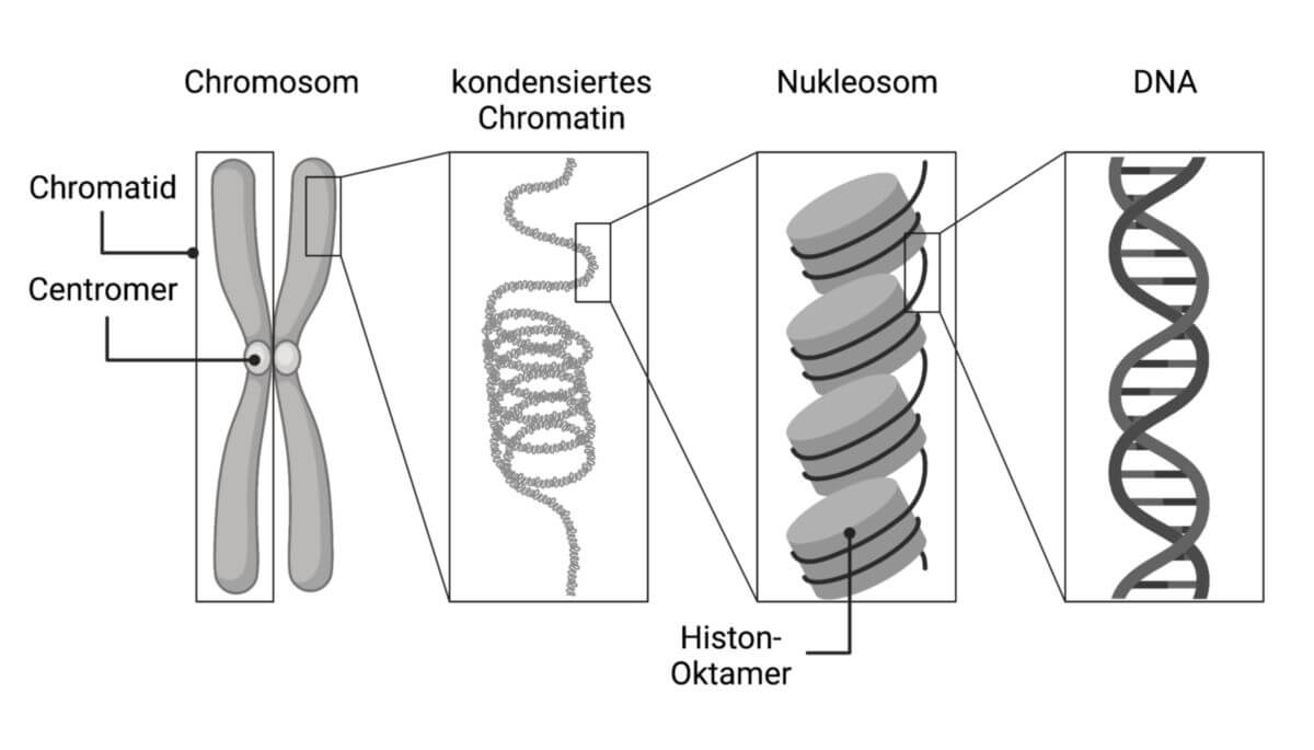
Chromosomen sind komplexe Strukturen im Zellkern von Eukaryoten, die aus DNA und Proteinen bestehen. Sie enthalten genetische Informationen der Zelle. Die chromosomale DNA formt durch Interaktion mit Histonen die Nukleosomen und wird dadurch komprimiert. Während der Kernteilung verdichten sich die Chromosomen und sehen stäbchenförmig aus. Die Gesamtheit des chromosomalen Materials wird als Chromatin bezeichnet.
Geschichte
Die Chromosomen wurden 1842 von Carl Wilhelm von Nägeli entdeckt. Thomas Hunt Morgan konnte 1910 in Drosophila melanogaster zeigen, dass die Chromosomen die Träger der Gene sind.
siehe auch: Chromosomentheorie der Vererbung
Aufbau
Allgemeine Organisation
Ein Chromosom besteht aus einem DNA-Doppelstrang, Histonen und Nicht-Histon-Proteinen. Während der Interphase befinden sich die Chromosomen im "entspannten" (entspiralisierten) Zustand als längere DNA-Fäden im Zellkern. Die DNA ist dabei in Abständen immer wieder um Proteinkomplexe aus Histonen gewickelt (Nukleosom). Dies dient der räumlichen Komprimierung. Die sich wiederholenden Nukleosomen ähneln optisch einer Perlenkette.
Auch im entspiralisierten Zustand unterscheidet sich der Packungszustand der DNA. So wird das Chromatin in zwei Formen aufgeteilt: locker gepacktes Euchromatin und dicht gepacktes Heterochromatin. Euchromatin enthält aktive DNA, die zugänglich für Enzyme ist und transkribiert wird. Heterochromatin enthält inaktive DNA, die nicht transkribiert wird.
Die Enden der Chromosomen werden als Telomere bezeichnet. An ihnen wird die DNA bei jeder Zellteilung ein wenig verkürzt. Telomere spielen daher bei Alterungsprozessen eine wichtige Rolle.
Während der Kernteilung beginnen sich die Chromosomen in der Prophase zu verdichten. Bei dieser Kondensation bilden sich aus den DNA-Fäden, durch mehrfache Schleifenbildung und Spiralisierung, stäbchenförmige Strukturen. Diese sind etwa 1 bis 50 µm lang, was ca. 1/10.000 der ursprünglichen Länge entspricht. Wie dieser Vorgang organisiert wird, ist derzeit (2023) noch unklar. Die resultierende Form wird als Metaphasenchromosom bezeichnet.
Metaphasenchromosom
Das Metaphasenchromosom ist die Form, auf die sich allgemein bezogen wird, wenn man von Chromosomen spricht. Nur in diesem kondensierten Zustand sind Chromosomen unter dem Lichtmikroskop erkennbar.
In der Metaphase bestehen Chromosomen aus zwei identischen Längseinheiten, den Chromatiden (Zwei-Chromatid-Chromosom). Sie werden auch als Schwesterchromatiden bezeichnet und enthalten ein identisches DNA-Molekül. Die Chromatiden sind durch das Centromer miteinander verbunden. Abhängig von der Lage des Centromers weisen sie eher eine X- oder V-Form auf. Es wird unterschieden zwischen:
- metazentrische Chromosomen Centromer in der Mitte
- akrozentrische Chromosomen Centromer zwischen Mitte und Telomer
- telozentrische Chromosomen Centromer in der Nähe des Telomers

In Ausnahmefällen treten auch dizentrische Chromosomen mit zwei Centromeren auf.
Neben der primären Einschnürungsstelle am Centromer können auch sekundäre Einschnürungen auftreten. Derart abgetrennte Bereiche des Chromosoms werden als Satelliten bezeichnet. Sie kodieren die ribosomale RNA und bilden die Nucleolus-Organisator-Region.
Chromosomenorganisation
Chromosomen sind hochgradig strukturiert. So liegen Gene mit ähnlicher Funktion im Chromosom häufig nebeneinander, obwohl sie auf dem linearen DNA-Strang an weit auseinanderliegenden Positionen kodiert sind.
Chromosomenbanden
Durch Anfärbung mit Giemsa-Lösung oder Quinacrin können die Chromosomen anhand ihres individuellen Bandenmusters unterschieden werden. Für die Giemsa-Färbung ist dafür zuvor eine Behandlung mit Trypsin notwendig. Es entstehen gefärbte G- (für Giemsa) und ungefärbte R-Banden (für Revers).
Das Färbeverhalten hängt mit der Basenverteilung zusammen. G-Banden haben einen höheren AT-Gehalt und enthalten wenige Gene, während R-Banden einen höheren GC-Gehalt haben und viele Gene aufweisen.
Nomenklatur
Für den Menschen und einige andere Modellorganismen existiert eine standardisierte Nomenklatur zur Bezeichnung der chromosomalen Regionen.[1] Dabei spielt die Lage des Centromers eine Rolle, die das Chromosom bzw. Chromatid in zwei Arme aufteilt: den kürzeren p-Arm (franz. petit = klein) und den längeren q-Arm. Die Banden werden vom Centromer ausgehend zum Telomer hin durchnummeriert und sind weiter in Sub-Banden unterteilt.
Beispiel: p53 ist kodiert am Genlokus 17p13.1, also auf dem kurzen Arm von Chromosom 17 in Bande 1, Sub-Bande 3 und Sub-Sub-Bande 1.
Das Bandenmuster aller menschlichen Chromosomen, inklusive der entsprechenden Benennung, sind auf der Ensembl-Website einsehbar.[2]
Kernteilung
Während der Kernteilung ist die chromosomale DNA nicht für Transkriptionsfaktoren oder Replikationsenzyme zugänglich, sondern liegt als inaktive Transportform vor.
Im Folgenden wird kurz die Teilung der Chromosomen während der Mitose und Meiose erläutert.
Mitose
- Prophase: Das Chromatin kondensiert zunehmend. Die Kernmembran wird aufgelöst.
- Metaphase: Die Chromosomen liegen als kondensierte Zwei-Chromatid-Chromosomen vor. Gegen Ende der Metaphase ordnen sie sich in der Äquatorialebene der Zelle an.
- Anaphase: Die Schwesterchromatiden werden am Centromer mithilfe des Spindelapparates getrennt. Die daraus entstandenen Ein-Chromatid-Chromosomen werden zu den entgegengesetzten Zellpolen gezogen.
- Telophase: Die Chromatiden entspiralisieren sich. Eine neue Kernmembran formt sich in jeder Tochterzelle.
Während der Interphase verdoppelt sich die DNA durch Replikation.
Meiose
Bei der Meiose finden zwei aufeinanderfolgende Zellteilungen statt, erst die Reduktions- und dann die Äquationsteilung. Bei beiden Teilungen kondensieren die Chromosomen, ähnlich wie bei der Mitose, während Pro- und Metaphase zu Zwei-Chromatid-Chromosomen.
Während der Reduktionsteilung findet die Paarung der homologen Chromosomen statt, bei dem sich mütterliches und väterliches Chromosom zusammenlagern und ein Bivalent bilden. Diese Phase der Prophase bezeichnet man als Zygotän. Im nachfolgenden Pachytän kommt es zum Crossing-over, bei dem sich die Chromatiden überkreuzen und mütterliches und väterliches Erbgut ausgetauscht wird. In der Anaphase werden die homologen Chromosomen getrennt und zu den entgegengesetzten Zellpolen gezogen. Es entstehen zwei haploide Tochterzellen.
Die anschließende Äquationsteilung findet analog zur Mitose statt: die Zwei-Chromatid-Chromosomen werden am Centromer getrennt und zwei Tochterzellen mit einem haploiden Ein-Chromatid-Chromosomensatz entstehen.
Chromosomenzahl
Innerhalb einer biologischen Spezies ist die Chromosomenzahl in den meisten Fällen gleich. Alle Chromosomen eines Individuums gemeinsam werden auch als Karyotyp bezeichnet.
Organismen, die sich ausschließlich asexuell vermehren, haben einen Chromosomensatz, der in allen Körperzellen gleich ist. Bei Organismen mit geschlechtlicher Fortpflanzung variiert die Chromosomenzahl in den Zellen und es kommen haploide (einfacher Chromosomensatz) und diploide (doppelter Chromosomensatz) Zellen vor:
- Diplont (z.B. Mensch): somatische Zellen (normale Körperzellen) mit diploidem Chromosomensatz (d.h. mit je einem Chromosom von den beiden Elternorganismen) sowie haploide Keimzellen. Diese verschmelzen bei der Befruchtung zur diploiden Zygote.
- Haplont (z.B. Pilze): Körperzellen sind haploid und verschmelzen zur diploiden Zygote. Die Zygote vollzieht eine Meiose und haploide Tochterzellen entstehen.
- Diplohaplont (z.B. Algen): Generationswechsel zwischen haploiden und diploiden Generationen
Bei manchen Spezies (u.a. bei Pflanzen und bestimmten Fischarten) können auch polyploide Chromosomensätze vorkommen.
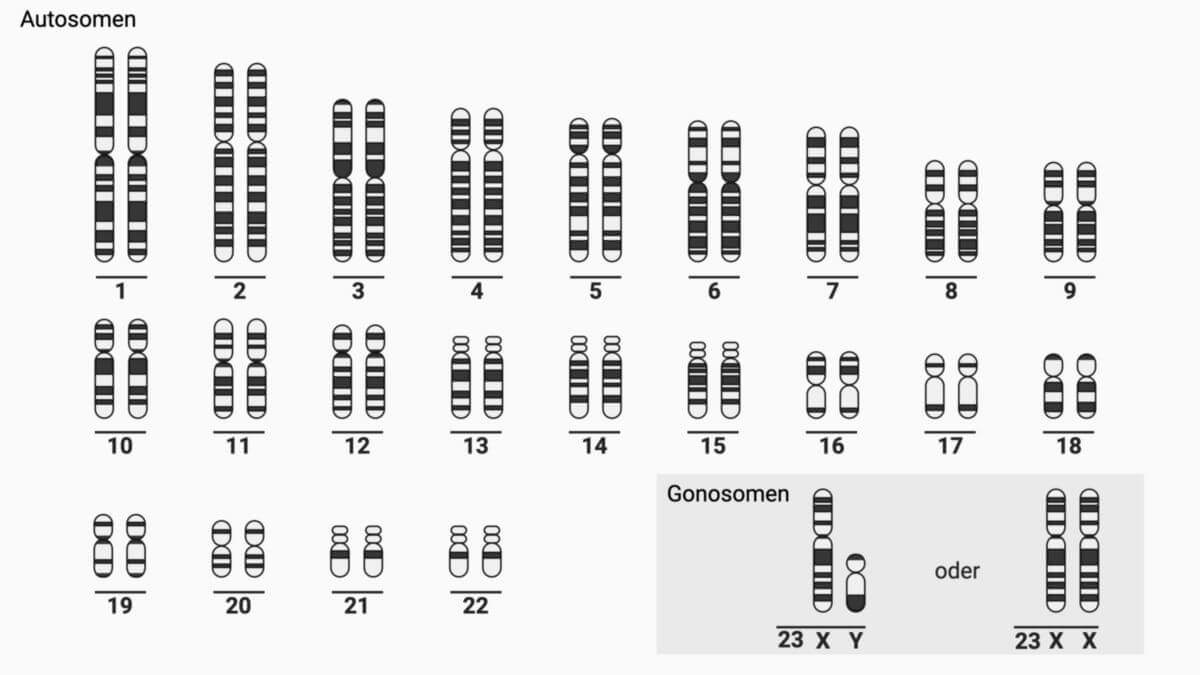
Menschliche Chromosomen
Der Mensch verfügt wie viele andere Organismen, die sich geschlechtlich fortpflanzen, über sogenannte Gonosomen. Diese sind spezielle Geschlechtschromosomen, die das biologische Geschlecht des Menschen determinieren. Frauen haben zwei X-Chromosomen und Männer je ein X- und ein Y-Chromosom. Bei Frauen wird eines der X-Chromosomen inaktiviert und erscheint unter dem Mikroskop als Barr-Körperchen oder Geschlechts-Chromatin.
Insgesamt weist das gesunde menschliche Genom 46 Chromosomen auf, davon 44 Autosomen (22 homologe Chromsomenpaare) und 2 Gonosomen. Diese sind in der folgenden Tabelle aufgelistet.[3]
| Art | Name | Länge (bp) | Anzahl kodierender Gene |
|---|---|---|---|
| Autosom | Chromosom 1 | 248.956.422 | 2.061 |
| Chromosom 2 | 242.193.529 | 1.299 | |
| Chromosom 3 | 198.295.559 | 1.081 | |
| Chromosom 4 | 190.214.555 | 757 | |
| Chromosom 5 | 181.538.259 | 882 | |
| Chromosom 6 | 170.805.979 | 1.051 | |
| Chromosom 7 | 159.345.973 | 1.012 | |
| Chromosom 8 | 145.138.636 | 702 | |
| Chromosom 9 | 138.394.717 | 778 | |
| Chromosom 10 | 133.797.422 | 730 | |
| Chromosom 11 | 135.086.622 | 1.317 | |
| Chromosom 12 | 133.275.309 | 1.038 | |
| Chromosom 13 | 114.364.328 | 323 | |
| Chromosom 14 | 107.043.718 | 821 | |
| Chromosom 15 | 101.991.189 | 617 | |
| Chromosom 16 | 90.338.345 | 863 | |
| Chromosom 17 | 83.257.441 | 1.186 | |
| Chromosom 18 | 80.373.285 | 266 | |
| Chromosom 19 | 58.617.616 | 1.476 | |
| Chromosom 20 | 64.444.167 | 546 | |
| Chromosom 21 | 46.709.983 | 221 | |
| Chromosom 22 | 50.818.468 | 495 | |
| Gonosom | X-Chromosom | 156.040.895 | 859 |
| Y-Chromosom | 62.460.029 | 106 |
Karyogramm
Um die Anzahl der Chromosomen eines Lebewesens festzustellen, müssen sie zunächst während der Metaphase im Reagenzglas mit Colchizin arretiert werden, so dass sie nicht mehr zu den Zellpolen gezogen werden. Die Chromosomen können dann angefärbt (der Name Chromosom leitet sich von ihrer Anfärbbarkeit her), fotografiert und in einem Karyogramm angeordnet werden.
Chromosomenaberrationen
Ein Fehler bei der Chromosomentrennung oder beim Crossing-over kann zu schweren Krankheiten führen. Diese lassen sich in zwei Klassen einteilen, denen jeweils einige Beispiele zugeordnet sind.
- Chromosomenaberrationen oder teilweise Chromosomendysplasie, gewöhnlich das Resultat eines fehlerhaften Crossing-over.
- Cri-du-chat Syndrom, das durch Deletion des kurzen Arms von Chromosom 5 hervorgerufen wird. Betroffene stoßen hochfrequente Laute aus, die an eine Katze erinnern. Sie haben weit auseinanderliegende Augen, einen kleinen Kopf und Kiefer, und sind geistig zurückgeblieben.
- Wolf-Hirschhorn-Syndrom, hervorgerufen durch teilweise Deletion des kurzen Arms von Chromosom 4. Betroffene leiden unter schweren Wachstums- und geistigen Störungen.
- Fehlende oder zusätzliche Chromosomen als Resultat einer unvollständigen chromosomalen Segregation.
- Down-Syndrom oder Trisomie 21 (zusätzliches Chromosom 21). Symptome sind zurückgebliebene Muskelentwicklung, asymmetrischer Schädel und geistige Retardierung.
- Klinefelter-Syndrom (XXY). Männer mit diesem Syndrom sind gewöhnlich steril, sehr groß, haben ungewöhnlich lange Arme und Beine, eine Tendenz zur Ausbildung von Brüsten und eine reduzierte Körperbehaarung. Weitere Symptome sind Emotionslosigkeit, Müdigkeit, Apathie und eine erhöhte Tendenz zu psychiatrischen Störungen. Eine verminderte Intelligenz, wie oft behauptet wird, scheint nicht zu bestehen.
- Turner-Syndrom (X0). Frauen mit diesem Syndrom haben unterentwickelte weibliche Geschlechtsmerkmale. Sie haben eine kleine Statur, einen tiefen Haaransatz, eine ungewöhnliche Augen- und Knochenentwicklung und eine Trichterbrust.
Diagnostik
Mittels Chromosomenanalyse können numerische und strukturelle Chromosomenaberrationen zytogenetisch nachgewiesen werden.
siehe auch: Geschlechtschromosom, Chromosomenmutation, Genom, Genommutation
Quellen
- ↑ Cytogenetic Banding Nomenclature, abgerufen am 12.11.2021
- ↑ Ensembl - Chromosomenbanden Mensch, abgerufen am 12.11.2021
- ↑ Ensembl - Menschliche Chromosomen Summary, abgerufen am 01.09.2023
Literatur
- Friedhelm Göltenboth: Chromosomenpraktikum. Stuttgart: Thieme-Verlag, 1978
- Walter Nagl: Chromosomen: Organisation, Funktion und Evolution des Chromatins. 2., neubearb. u. erw. Aufl. Berlin: Parey-Verlag 1980
- Rigomar Rieger: Struktur und Strukturumbauten der Chromosomen vielzelliger Organismen. Berlin: Akademie-Verlag 1979
- Gholamali Tariverdian: Chromosomen, Gene, Mutationen: humangenetische Sprechstunde. Berlin: Springer-Verlag 1995
- Walther Traut: Chromosomen: klassische und molekulare Cytogenetik. - Berlin: Springer-Verlag 1991