Calcaneus


von lateinisch: calcar - Sporn
Synonym: Fersenbein, Os calcis
Englisch: calcaneus
Definition
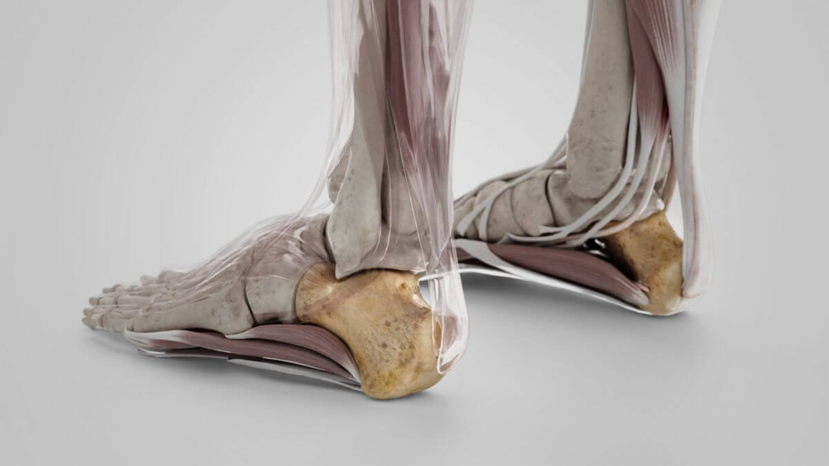
Der Calcaneus ist der größte Knochen des Fußskeletts. Er ist an der Bildung des unteren Sprunggelenks (USG) beteiligt und dient als Hebelarm für die Flexoren des Unterschenkels, die über die Achillessehne an ihm ansetzen.
Anatomie
Der Calcaneus hat eine würfelförmige Grundform und weist 6 Flächen auf.
Flächen
Facies superior
Die Facies superior des Calcaneus wird im Wesentlichen von 3 Gelenkflächen eingenommen, die mit dem Talus artikulieren:
- Facies articularis talaris posterior
- Facies articularis talaris media
- Facies articularis talaris anterior
Zwischen der Facies articularis talaris posterior und media liegt der Sulcus calcanei. Er formt gemeinsam mit dem Sulcus talaris des Talus den so genannten Sinus tarsi, einen Tunnel, der das Ligamentum talocalcaneum interosseum aufnimmt. Die Facies articularis talaris posterior ist an der Bildung des hinteren USG (Articulatio subtalaris) beteiligt, die Facies anterior und Facies media an der Bildung des vorderen USG (Articulatio talocalcaneonavicularis). Die kleine, raue Knochenfläche vor den Gelenkflächen dient als Ursprung des Musculus extensor digitorum brevis.
Facies inferior (Plantarfläche)
Die Facies inferior ist uneben und hinten breiter als vorne. Sie wird posterior von einer prominenten Erhebung, dem Fersenhöcker (Tuber calcanei), begrenzt. An ihm setzt die Achillessehne an. Das Tuber bildet an beiden Seiten jeweils einen kleinen Knochenfortsatz (Processus) aus.
Der zur Innenseite hin gelegene Processus medialis (tuberis calcanei) dient als Ursprung für den Musculus abductor hallucis und den Musculus flexor digitorum brevis. Von seiner Basis bis zum Processus lateralis (tuberis calcanei), der an der Außenseite des Fußes gelegen ist, läuft die Ursprungsfläche des Musculus abductor digiti quinti quer über den Knochen. Die beiden Processus dienen darüber hinaus der Verankerung der Plantaraponeurose.
Zwischen den beiden Processus befindet sich eine zentral verlaufende Einsenkung. Weiter anterior vor den Processus setzen medial und lateral die beiden Köpfe des Musculus quadratus plantae an.
Facies medialis
Die mediale Seite des Calcaneus ist konkav ausgeformt. An ihrem kranialen Ende befindet sich ein markanter, horizontal verlaufender Knochenvorsprung, das Sustentaculum tali. Die Unterseite ist rinnenförmig zum Sulcus tendinis musculi flexoris hallucis longi ausgeformt und nimmt die Sehne des Musculus flexor hallucis longus auf.
Facies lateralis
Die laterale Seite des Calcaneus hat einen leicht vorgewölbten Knochenhöcker (Trochlea fibularis). Unter der Trochlea fibularis liegt der Sulcus tendinis musculi peronei longi, der die Sehne des Musculus peroneus longus aufnimmt.
Facies anterior
Die vordere Stirnseite des Calcaneus trägt mit der Facies articularis cuboidea eine Verbindungsfläche zum Os cuboideum.
Entwicklung
Der Knochenkern des Calcaneus entwickelt sich zwischen dem 4. und 7. Fetalmonat.
Funktion
Der Calcaneus dient als Hebelarm für die wichtigsten Flexoren der Unterschenkelmuskulatur. Ferner hat er durch den Ursprung der Plantaraponeurose einen wesentlichen Anteil an der Aufrechterhaltung der Spannung des Fußgewölbes.
Klinik
Häufige pathologische Veränderungen des Calcaneus sind der sogenannte Fersensporn und die Haglund-Deformität. Dabei kommt es zu einer Verknöcherung der Sehnenansätze der Plantaraponeurose ("plantarer Fersensporn") bzw. der Achillessehne ("kranialer Fersensporn"). Beide Varianten bleiben häufig asymptomatisch, können aber auch zu Belastungsschmerzen führen.
Kalkaneusfrakturen sind aufgrund der kompakten Bauweise des Knochens eher selten und treten vor allem bei massiver Gewalteinwirkung auf, z.B. beim Fall aus großer Höhe. Ermüdungsfrakturen des Calcaneus kommen bei Sportlern vor, die Sprungsportarten betreiben.