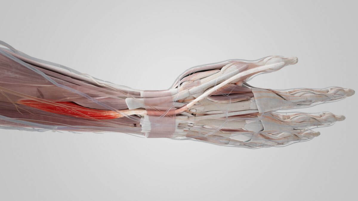
Corpus: Extensor pollicis longus muscle

from latin: pollex - thumb
Definition
Anatomy
Origin
The origin of the muscle is on the posterior surface of the ulna and the interosseous membrane of the forearm, close to the origins of the abductor pollicis longus and the extensor pollicis brevis muscles.
Insertion
The extensor pollicis longus muscle attaches to the dorsal side of the distal phalanx of the thumb.
Innervation
The innervation is provided by the posterior interosseous nerve from the deep branch of the radial nerve with fibers from the C7 and C8 segments.
Blood Supply
The extensor pollicis longus muscle and its tendon receive blood from various arteries. The muscle belly is supplied by branches of the posterior interosseous artery. The tendon is supplied proximally by branches of the anterior interosseous artery and distally by branches of the radial artery and the dorsal metacarpal artery I.
Topography
The tendon of the extensor pollicis longus muscle runs distally on the dorsal side of the radius. It lies in a tight, obliquely running groove at the end of the radius and passes through the third tendon compartment of the extensor retinaculum. On its path, it uses the Lister's tubercle as a pulley. Along with the tendons of the extensor brevis muscle and the abductor pollicis longus muscle, the insertion tendon of the extensor pollicis longus muscle crosses the radial artery. It limits the anatomical snuffbox (Fovea radialis).
Function
The extensor pollicis longus is responsible for the extension and abduction of the thumb. Its action is diminished by an extension of the hand at the wrist.
Clinic
The tendon of the muscle is visible from the outside as it forms the anterior boundary of the snuffbox. A tear of the tendon leads to so-called "drummer's palsy." Reconstruction can be performed through an indicisplasty.